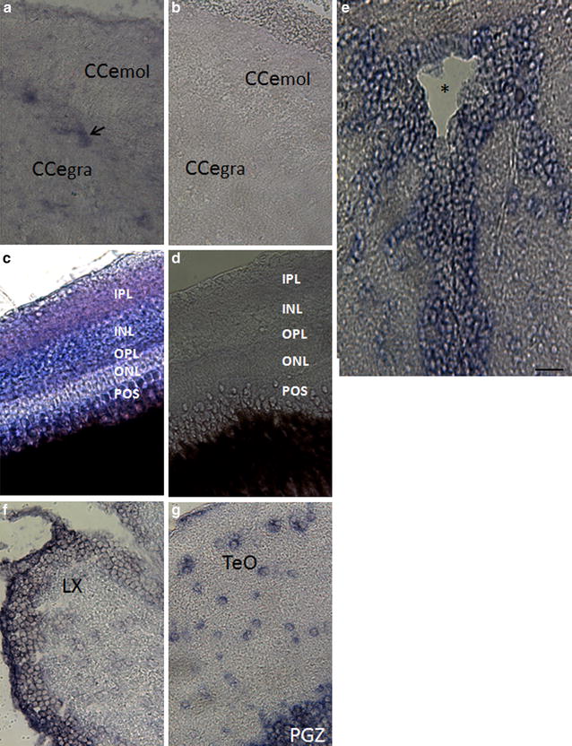
Fig. 7 wdr81 expression in the adult brain and eye tissues. wdr81 expression was detected in the cerebellum (a), retina (c), tectal ventricle (e), brain stem (f), and optic tectum (g). Results with a sense probe in both cerebellum (b) and retina (d), which demonstrate no staining, indicates the specificity of the signal obtained with an antisense probe and both cerebellum (b) and retina (d) are shown. CCe mol Cerebellar molecular layer, Cce gra cerebellar granular layer, POS photoreceptor outer segments, ONL outer nuclear layer, OPL outer plexiform layer, INL inner nuclear layer, IPL inner plexiform layer, LX lobus vagus, TeO optic tectum, PGZ periventricular gray zone of the optic tectum. The arrow indicates the Purkinje cell layer and the asterisk the tectal ventricle. Scale bar equals 200 μm
Image
Figure Caption
Figure Data
Acknowledgments
This image is the copyrighted work of the attributed author or publisher, and
ZFIN has permission only to display this image to its users.
Additional permissions should be obtained from the applicable author or publisher of the image.
Full text @ BMC Neurosci.

