Image
Figure Caption
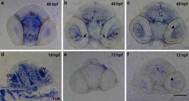
Fig. 5 Transverse sections through the head regions of whole mount in situ hybridization specimens at 3 embryonic timepoints. The expression of wdr81 was observed in a regionally-specific manner by 48 hpf (a–c), whereas it was ubiquitously expressed at 18 hpf (d), and decreased by 72 hpf (e, f). Arrows indicate the optic nerve, asterisk the region of the nucleus of the medial longitudinal fascicle, and arrowhead the retina. Po preoptic area, Di diencephalon, T. midbrain tegmentum, Le lens, OV optic vesicle, Yolk yolk sac. Scale bar equals 100 μm
Figure Data
Acknowledgments
This image is the copyrighted work of the attributed author or publisher, and
ZFIN has permission only to display this image to its users.
Additional permissions should be obtained from the applicable author or publisher of the image.
Full text @ BMC Neurosci.

