Image
Figure Caption
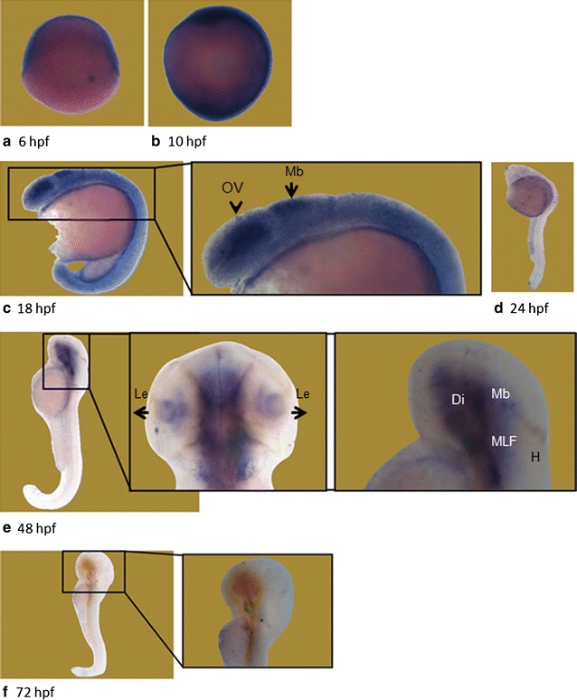
Fig. 4 Whole mount in situ hybridization revealed differential expression of wdr81 transcript during embryonic development. Our results from the WMISH method are in parallel with the qRT-PCR data. The signal is high during the first 3 developmental timepoints (6–18 hpf), it is decreased and maintained during the rest of the development periods (24–72 hpf). OV optic vesicle, Mb midbrain, Le lens, H hindbrain, Di diencephalon, MLF medial longitudinal fascicle.
Figure Data
Acknowledgments
This image is the copyrighted work of the attributed author or publisher, and
ZFIN has permission only to display this image to its users.
Additional permissions should be obtained from the applicable author or publisher of the image.
Full text @ BMC Neurosci.

