Image
Figure Caption
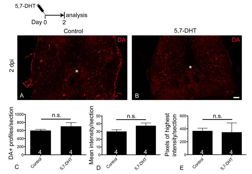
Fig. S3 Spinal dopamine abundance is not detectably altered after ablation of serotonergic axons with 5,7-DHT.
A,B: Cross sections through the spinal cord are shown; asterisks indicate the central canal. Immunohistochemistry for dopamine reveals dopaminergic axons in the spinal cord of vehicle-injected and 5,7-DHT injected animals at 2 days post-injection.
C-E: Number of axonal profiles (Student’s t-test, P = 0.3787), as well as mean (P = 0.1875) and peak (P = 0.4724) intensities of dopamine labeling were not detectably different between control and toxin treated animals.
Scale bar in B = 50 µm.
Acknowledgments
This image is the copyrighted work of the attributed author or publisher, and
ZFIN has permission only to display this image to its users.
Additional permissions should be obtained from the applicable author or publisher of the image.
Full text @ Cell Rep.

