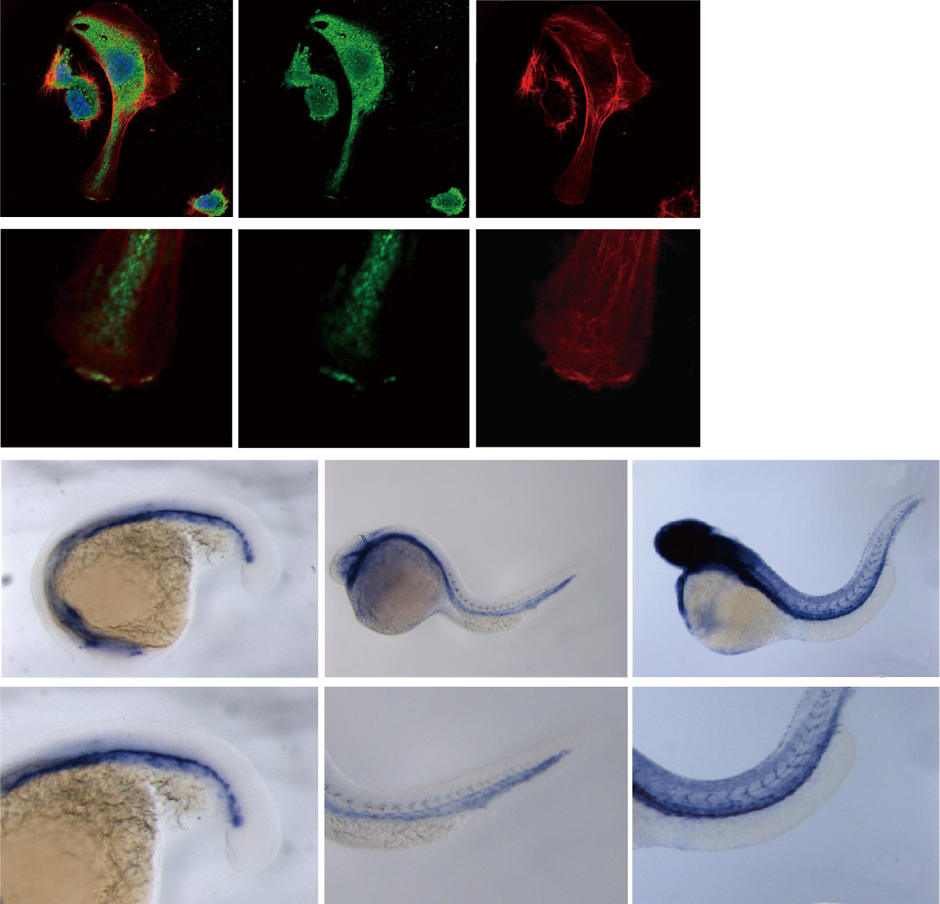
Fig. 1
PLK2 is expressed in endothelial cells.(A?B′′) Immunostaining of HUVECs reveals that PLK2 is expressed in ECs. (B?B′′) Enlarged image of the boxed area in A?A′′ shows that PLK2 can aggregate at the leading edge of extending ECs (arrowhead). (A and B)-merge; (A′ and B′)-anti-PLK2 immunostaining (green); (A′′ and B′′)-phalloidin actin staining (red). DAPI nuclear staining (blue). Scale bar, 40 µm. (C) Quantitative measurements of PLK2 localized at the leading edge and in the cytoplasm of HUVECs (Meanħs.e.m. *p=0.0056 by Student′s t-test). (D?F) in situ hybridization shows that plk2b is expressed in the zebrafish vasculature at 16 hpf (n=31/31), 24 hpf (n=46/47), and 48 hpf (n=28/30). (D′?F′) Enlarged image of the boxed area of D?F shows that plk2b is expressed in the cardinal vein, aorta, and intersomitic vessels of the zebrafish body and tail.
Reprinted from Developmental Biology, 404(2), Yang, H., Fang, L., Zhan, R., Hegarty, J.M., Ren, J., Hsiai, T.K., Gleeson, J.G., Miller, Y.I., Trejo, J., Chi, N.C., Polo-like kinase 2 regulates angiogenic sprouting and blood vessel development, 49-60, Copyright (2015) with permission from Elsevier. Full text @ Dev. Biol.

