
|
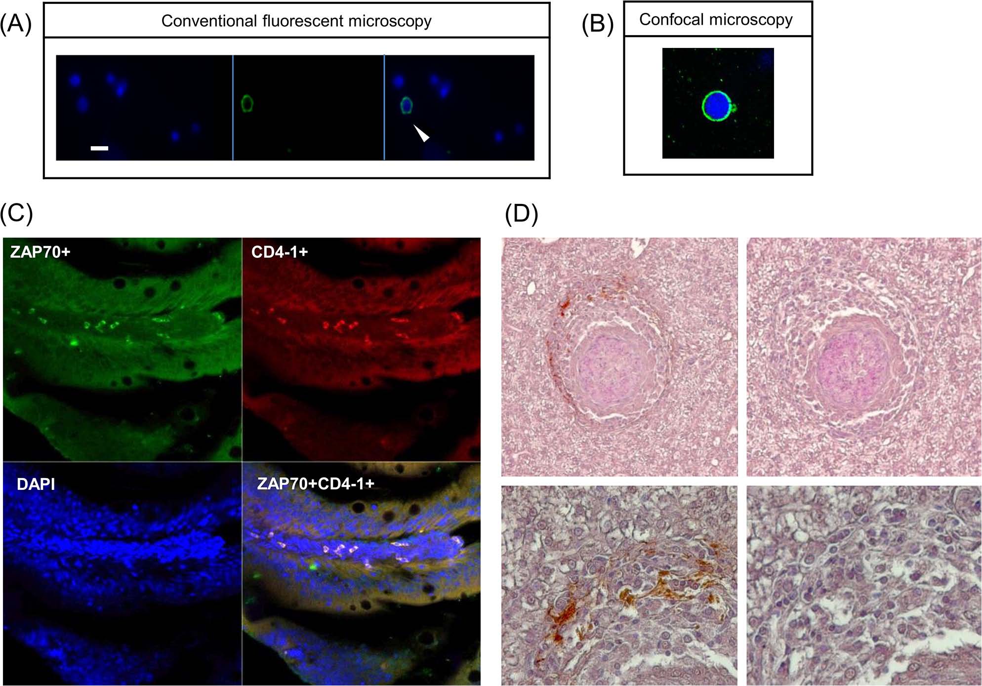
Fig. 7 Detection of zfCD4-1+ cells using immunofluorescence assay and immuohistochemical assays.
(A) Immunofluorescence staining of the zfCD4-1 molecule on zebrafish leukocytes. The cells stained with DAPI (blue) for counterstaining nuclei (left panel), the zfCD4-1 polyclonal followed by FITC labelled (green) secondary antibody (middle panel) and merged image (right panel) are shown. Scale bar = 10 �m). The arrow shows a cell expressing CD4 on its surface. (B) Cells from (A) were also visualized using a confocal microscope. (C) Double immunofluorescence staining of zebrafish peripheral lymphocytes in gut sections incubated with rabbit anti zfCD4-1 and mouse anti human ZAP70. ZAP70+ cells are green and CD4+ cells are red. Co-localization was confirmed by a Z-stack image analysis using Zeiss confocal microscopy. The data are representative results obtained from three independent experiments. (D) CD4-1+ cells present within the cuff of leukocytes surrounding the granulomas developed after M. marinum EspG5::Tn mutant infection of zebrafish 4 weeks earlier. Top images 200x, bottom images 400x. Left hand images used anti-CD4-1 serum, right hand images are controls.