
|
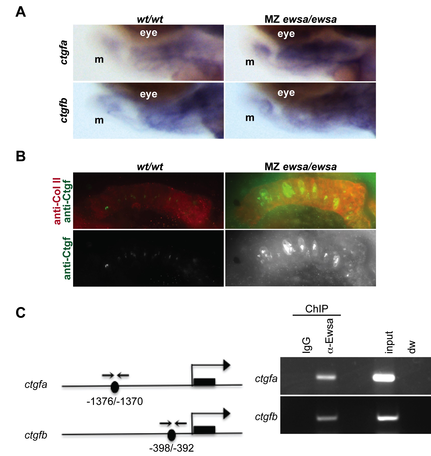
Fig. 7
Ewsa binds to ctgfa and ctgfb target genes.
A. Lateral views (anterior to the left, dorsal to the top) of 4dpf of (Left) wt/wt and (Right) MZ ewsa/ewsa visualized by in situ hybridization using probe for ctgfa (tom panel) and ctgfb (bottom panel). m: Meckel′s cartilage. B. Ventral views (anterior to the left) of 4dpf Meckel′s cartilage of (Left) wt/wt and (Right) MZ ewsa/ewsa visualized by immunohistochemistry using anti-Ctgf antibody (green), and anti-Collagen type II antibody (Red). C. (left) Schematics of ctgfa and ctgfb genes. Black circle: Sox9 binding site, black square: exon, arrows: PCR primer for ChIP assay. (right) ChIP assays were performed using 27 hpf zebrafish embryos. IgG: negative control of ChIP, anti-Ewsa: ChIP sample using EWSa antibody, input: 4.5% of DNA from total lysates was subjected to PCR, dw: negative control for PCR reaction.