
|
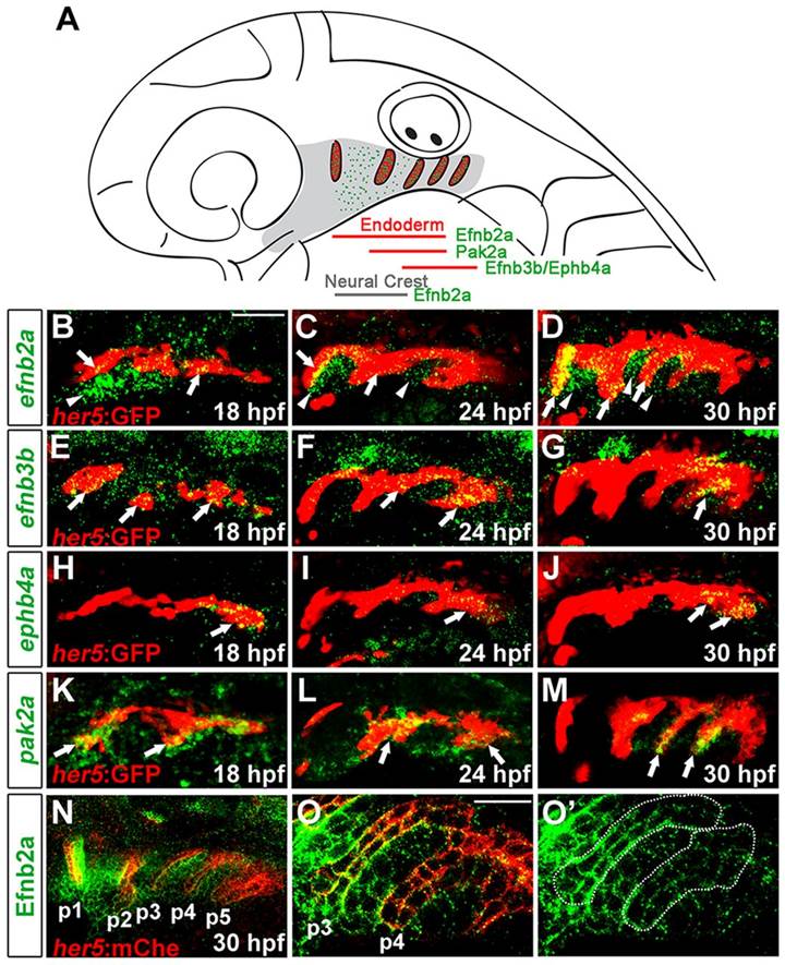
Fig. 1
Eph-ephrin and pak2a expression in pouches. (A) Schematic of pouches and expression of efnb2a, efnb3b, ephb4a and pak2a within the 30hpf zebrafish head. (B-M) Fluorescent in situ hybridization (green) shows a time-course of expression for efnb2a, efnb3b, ephb4a and pak2a relative to her5:GFP+ endodermal pouches (labeled red by GFP immunohistochemistry). Arrows show expression in pouch endoderm and arrowheads show expression in neural-crest-derived cells. Scale bar: 40�m. (N-O′) Anti-Efnb2a staining (green) relative to her5:mCherryCAAX+ endoderm (red) shows increased expression in more mature anterior pouches (e.g. p3) relative to less mature posterior ones (p4 and p5), as well as in neural-crest-derived cells. Dotted lines in O′ denote pouch expression. Scale bar: 20�m.