Fig. S1
Characterization of Stable Runx:GFP and Runx:mCherry Transgenic Zebrafish Lines, Related to Figure 1
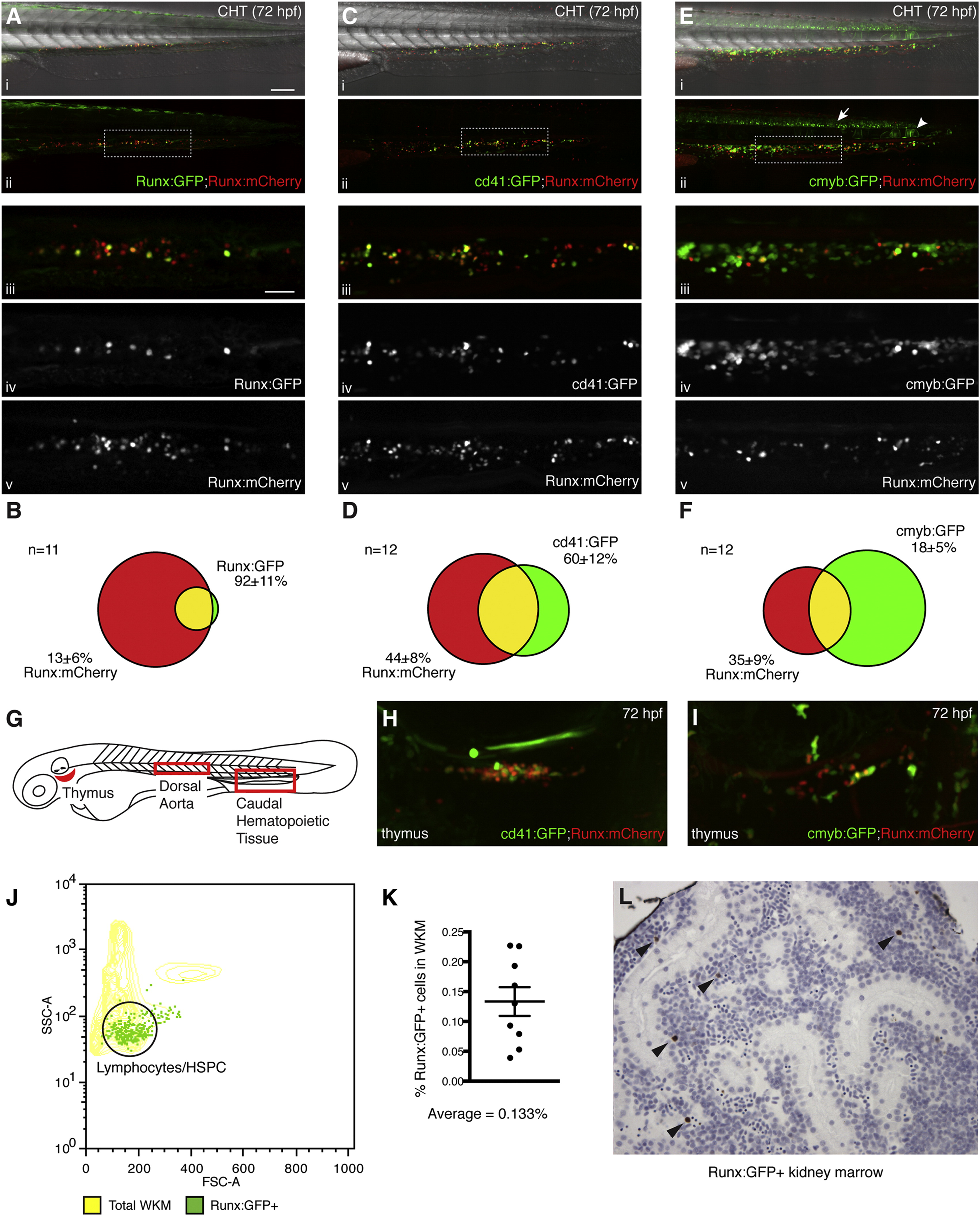
(A?F) Intercrosses of transgenic lines to show overlapping populations. Wide-field view of CHT at 72 hr postfertilization (hpf) with merged (i) GFP, mCherry and DIC channels, or (ii) GFP and mCherry. Detail in (ii) is shown below as (iii) merged GFP and mCherry, (iv) GFP, or (v) mCherry. Anterior left, posterior right, dorsal up, ventral down. Scale bars: (i-ii) 100 Ám, (iii-v) 30 Ám.
(A) Overlap of Runx:GFP and Runx:mCherry lines.
(B) Quantification of Runx:GFP and Runx:mCherry overlap. Runx:GFP overlaps 92 ▒ 11% with Runx:mCherry. Runx:mCherry overlaps 13 ▒ 6% with Runx:GFP. (CHT of n = 11 embryos scored).
(C) Overlap of cd41:GFP and Runx:mCherry lines.
(D) Quantification of cd41:GFP and Runx:mCherry overlap. cd41:GFP overlaps 60 ▒ 12% with Runx:mCherry. Runx:mCherry overlaps 44 ▒ 8% with cd41:GFP. (CHT of n = 12 embryos scored). cd41:gfp has known broader expression in thrombocyte progenitors at this stage (Bertrand et al., 2008 and Lin et al., 2005).
(E) Overlap of cmyb:GFP and Runx:mCherry lines. (ii) Note the expression of the cmyb:GFP transgene in the ventral neural tube (arrow) and posterior notochord (arrowhead).
(F) Quantification of cmyb:GFP and Runx:mCherry overlap. cmyb:GFP overlaps 18 ▒ 5% with Runx:mCherry. Runx:mCherry overlaps 35 ▒ 9% with cmyb:GFP. (CHT of n = 12 embryos scored).
(G) Schematic of 60 hpf zebrafish embryo with location of thymus, dorsal aorta and caudal hematopoietic tissue (CHT).
(H) Overlap of cd41:GFP and Runx:mCherry lines in the developing thymus. Note the bright circulating thrombocyte above.
(I) Overlap of cmyb:GFP and Runx:mCherry lines in the developing thymus.
(J) Forward scatter (FSC) and side scatter (SSC) were used to distinguish characteristic whole kidney marrow (WKM) populations (Traver et al., 2003). Runx:GFP+ cells were found predominately in the lymphoid/HSPC gate.
(K) Flow cytometry of WKM from adult Runx:GFP transgenic zebrafish. Runx:GFP+ cells represent on average 0.133% of the total WKM.
(L) Immunohistochemistry with anti-GFP antibody shows rare GFP+ cells in sections of Runx:GFP kidney marrow (brown; arrowheads; counterstained with hematoxilyn).
Error bars show mean ▒ SEM.
Reprinted from Cell, 160, Tamplin, O.J., Durand, E.M., Carr, L.A., Childs, S.J., Hagedorn, E.J., Li, P., Yzaguirre, A.D., Speck, N.A., Zon, L.I., Hematopoietic Stem Cell Arrival Triggers Dynamic Remodeling of the Perivascular Niche, 241-252, Copyright (2015) with permission from Elsevier. Full text @ Cell

