
Fig. 7
|
Fig. 7
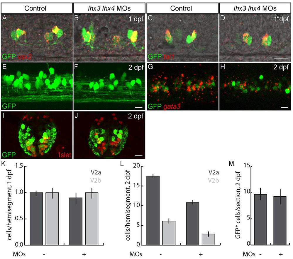
Lhx3 and Lhx4 promote differentiation of late-born V2a and V2b INs. (A-D) Lateral views of 24hpf Tg(vsx1:GFP) embryos. (A,B,K) The number of early-born V2a INs labeled with vsx2 is unchanged in lhx3+lhx4 MO-injected embryos. (C,D,K) The number of early-born V2b INs labeled with tal1 is unchanged in lhx3+lhx4 MO-injected embryos. (E-H) Lateral views of 2dpf Tg(vsx1:GFP) embryos. (E,F) V2as labeled by GFP in 2dpf Tg(vsx2:EGFP) embryos are reduced in lhx3+lhx4 MO-injected embryos. (G,H) Single optical section; the number of V2bs co-labeled by gata3 and GFP in Tg(vsx1:GFP) embryos is reduced in lhx3+lhx4 MO-injected embryos. (I,J) Cross-section through mid-trunk of 2dpf Tg(vsx1:GFP) embryos. The number of V2s and late p2 progenitors labeled by GFP is unaffected in lhx3+lhx4 MO-injected embryos. Islet was included to exclude MNs that weakly express GFP (Reimer et al., 2013). (K) Quantification of early-born V2a and V2b INs (P>0.05; n=40 segments in ten embryos for each condition). (L) Quantification of V2a and V2b INs at 2dpf (PV2a=1�1011; PV2b=2�105; n=60 segments from ten embryos for each condition). (M) Quantification of V2 INs plus late p2 progenitors (P=0.14; n=10 alternating segments from ten embryos for each condition). In control embryos some weakly GFP+ cells co-express Islet; these cells are MNs and were not included in p2 progenitor and V2 IN counts. Scale bars: 20�m.