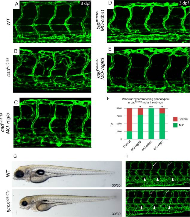
Fig. 3 cad artery hyperbranching is Vegfc/Vegfr3 dependent. A,B: The vasculature Tg(fli1a:EGFP) in wild-type sibling (A) and cadhu10125 (B) embryos at 3 dpf. C?E: The vasculature Tg(fli1a:EGFP) in cadhu10125/MO-vegfc (C), cadhu10125/MO-ccbe1 (D), and cadhu10125/MO-vegfr3 (E) embryos at 3 dpf. F: Quantification of the severity of vascular hyperbranching designated as mild or severe (where mild: 1?7 hyperbranched arteries, and severe: e 8 hyperbranched arteries along the entire embryo body axis) scored in WT (n = 16), MO-vegfc (n = 12), MO-ccbe1 (n = 10) and MO-vegfr3 (n = 14) injected cadhu10125 embryos. G,H: The thymidylate synthase mutant tymshi3510Tg does not present with arterial hyperbranching. G: Overall morphology of wild-type (n = 30/30) and tymshi3510Tg (n = 30/30) embryos at 4 dpf. H: The vasculature Tg(fli1a:EGFP) in wild-type (n = 30/30) and tymshi3510Tg (n = 30/30) embryos at 4dpf. White arrowhead: thoracic duct, white arrow: example of normal aISV.
Image
Figure Caption
Figure Data
Acknowledgments
This image is the copyrighted work of the attributed author or publisher, and
ZFIN has permission only to display this image to its users.
Additional permissions should be obtained from the applicable author or publisher of the image.
Full text @ Dev. Dyn.

