
|
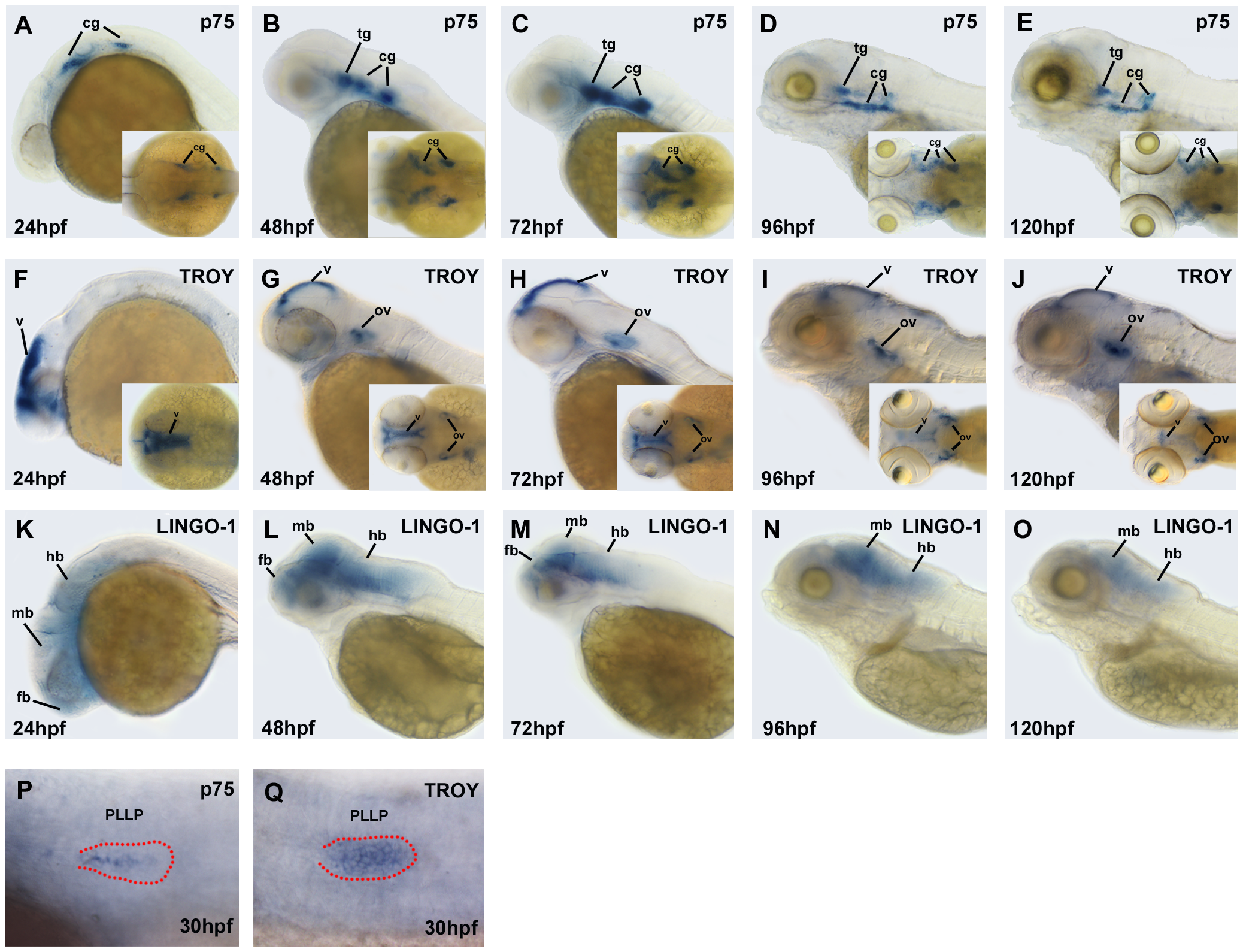
Fig. 1
Expression patterns of Nogo co-receptor mRNAs in zebrafish embryos.
Whole-mount in situ hybridization was performed with antisense probes against p75 (A?E; P), TROY (F?J; Q), and LINGO-1 (K?O) at the indicated developmental stages. Images were taken from the lateral view with the anterior to the left and the dorsal to the top, or from the dorsal view (inset). The PLL primordium is visible in the embryos with DIC optics, and labeled with red dots in panels (P) and (Q). fb, forebrain; mb, midbrain; hb, hindbrain; tg, trigeminal ganglion; cg, cranial ganglion; v, ventricle; ov, otic vesicle; PLLP, posterior lateral line primordium.