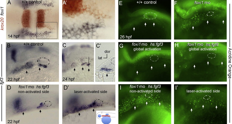
Fig. 9
Fgf mis-expression in zebrafish rescues the cell death phenotype in foxi1 morphants. (A) and (A2) At 14 hpf (10 somites), krox20 (red) marks streams of nascent neural crest migrating from the hindbrain whereas foxi1 (black) marks non-neural ectoderm abutting the hindbrain. No cells co-express both markers. The boxed area in (A) is magnified in (A2). Images show a dorsal view with anterior to the left. (B?I2) Embryos at later stages are oriented with anterior to the left and dorsal up, with the otic vesicle circled with a dashed line and pharyngeal pouches marked with arrows. (B?C2) Expression of fgf3 in wild-type control embryos marks pharyngeal pouch endoderm at 22 hpf (B) and 24 hpf (C and C2). The vertical line in (C) marks the plane of section in (C2). (D and D2) A hs:fgf3 transgenic embryo injected with foxi1-MO shows a reduced level of fgf3 expression on the non-activated side at 22 hpf (D) but shows dramatically elevated expression on the laser-activated side (D2). Note the horizontal axis in D is inverted to facilitate comparison. Laser-activation was performed at 20 hpf, focusing on the pharyngeal region, shown in the inset to panel (D2). (E?I2) Live embryos were incubated with acridine orange at 26 hpf to label cells undergoing apoptosis. A wild-type control embryo shows very few apoptotic cells (E) whereas a foxi1 morphant shows a marked increase in apoptosis (F). The cell death phenotype normally seen in foxi1 morphants was strongly suppressed by global low-level activation of hs:fgf3 (37 °C for 30 min beginning at 20 hpf) (G) or hs:fgf8 (35 °C for 6 h beginning at 20 hpf) (H). A hs:fgf3 transgenic embryo injected with foxi1-MO showed elevated apoptosis on the non-activated side ((I) horizontal axis inverted for easier comparison), but apoptosis was strongly suppressed on the laser-irradiated side (I2).
Reprinted from Developmental Biology, 390, Edlund, R.K., Ohyama, T., Kantarci, H., Riley, B.B., Groves, A.K., Foxi transcription factors promote pharyngeal arch development by regulating formation of FGF signaling centers, 1-13, Copyright (2014) with permission from Elsevier. Full text @ Dev. Biol.

