
|
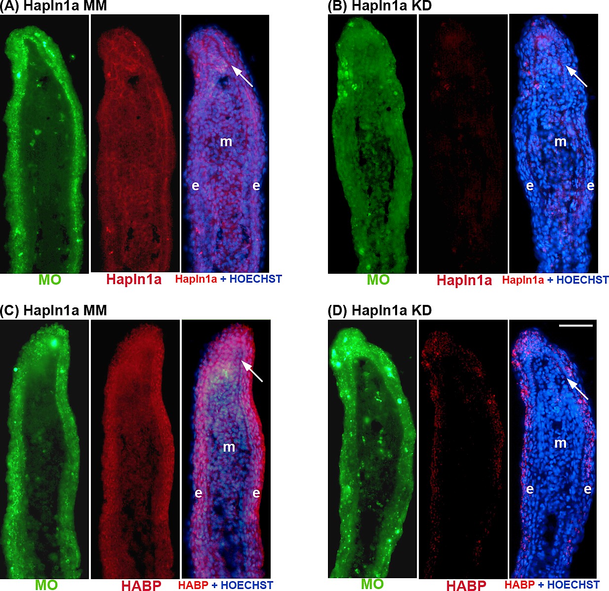
Fig. S2
The hapln1a-KD was effective in alf dty86. Immunostaining for Hapln1a or HA (Red) and HOECHST staining for DNA (blue). The green reveals the location of the targeting and control MOs, which are fluroescein tagged (A) Longitudinal section of an alf dty86 fin ray treated with Hapln1a control morpholino (MM). (B) Longitudinal section of an alf dty86 fin ray knocked down for Hapln1a with a targeting morpholino (KD). Compared to the control MM fins, Hapln1a knock-down (KD) fins exhibit reduced staining for Hapln1a. HA was detected by biotinylated-HABP followed by streptavidin-Alexa 546 conjugate. (C) Longitudinal section of an alf dty86 fin ray treated with Hapln1a control morpholino (MM). (D) Longitudinal section of an alf dty86 fin ray knocked down for Hapln1a with a targeting morpholino (KD). Compared to the control MM fins, Hapln1a knock-down (KD) fins exhibit reduced staining for HA. Arrow identifies the basal layer of the epidermis; m, mesenchyme; e, epithelium. Scale bar is 50 μm.