Fig. 2
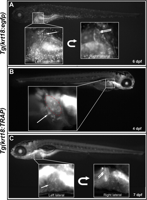
krt18-driven reporters allow live detection of the developing biliary system. (A) Fluorescent micrograph of live 6 dpf Tg(krt18:egfp) larva, showing EGFP expression in intrahepatic biliary cells (single arrow) and gallbladder epithelium (double arrow), partly obscured by skin expression. (B) and (C) Fluorescent micrographs of live 4 dpf (B) and 7 dpf (C) Tg(krt18:TRAP) larvae, showing visualization of EGFP-Rpl10a fusion protein in developing intrahepatic biliary cells (single arrows) and gallbladder epithelium (double arrow). Red dashed line in (B) indicates gallbladder overlying the liver, out of the focal plane.
Reprinted from Gene expression patterns : GEP, 14(2), Wilkins, B.J., Gong, W., and Pack, M., A novel keratin18 promoter that drives reporter gene expression in the intrahepatic and extrahepatic biliary system allows isolation of cell-type specific transcripts from zebrafish liver, 62-68, Copyright (2014) with permission from Elsevier. Full text @ Gene Expr. Patterns

