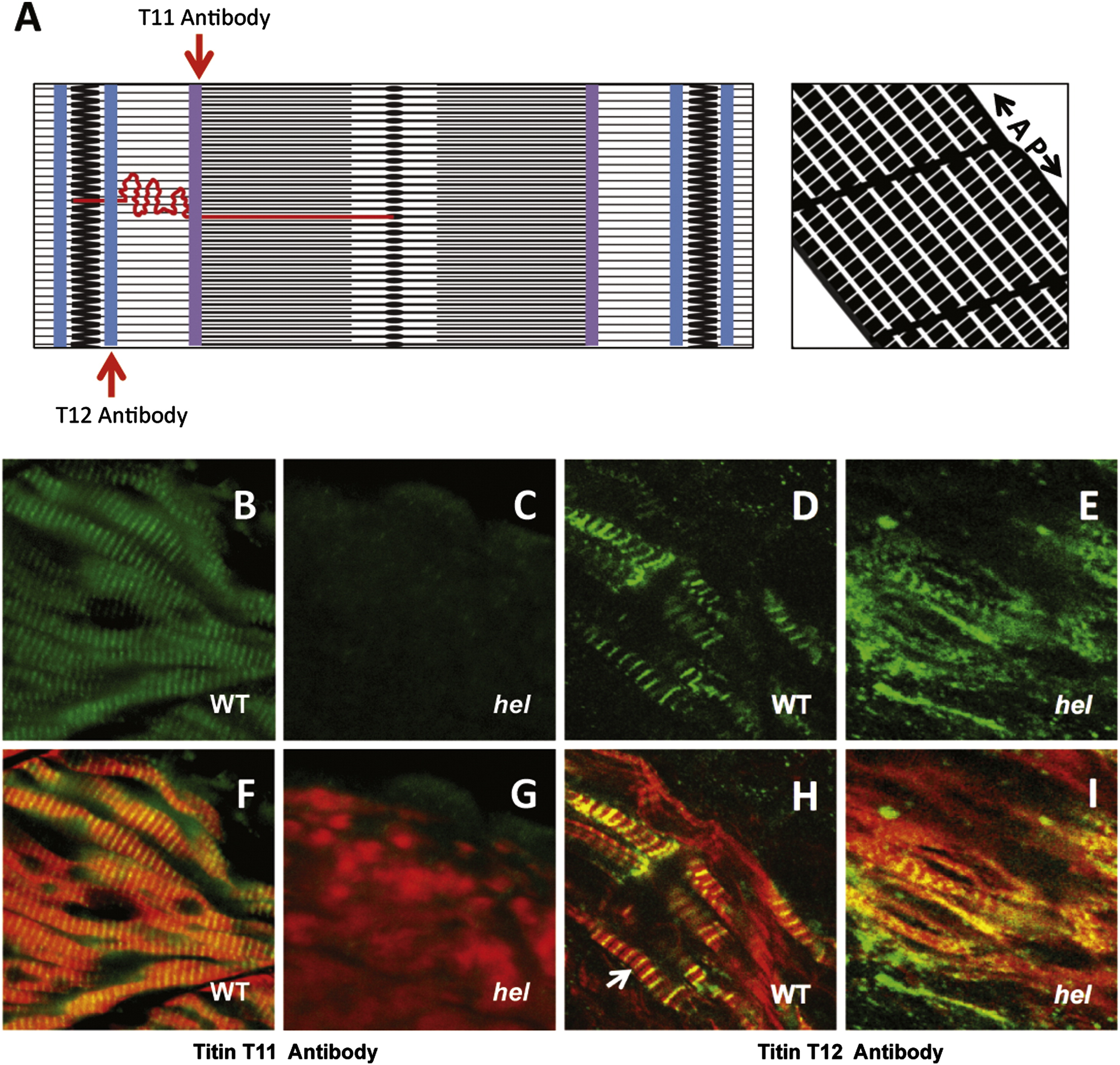
Fig. 4 The herzschlag mutation results in a form of titin that fails to cross-react with antibodies specific for the thick filament rod domain. Immunofluorescent staining with antibodies directed against specific peptides of the titin protein shows that mutant embryos lack the T11 peptide, found at the I-band to A-band transition, but retain the T12 peptide, which localizes just lateral to the Z-disk (represented schematically in panel A). A?P indicates the direction of the anterior?posterior axis. Whole-mount WT hel siblings (B, D, F, and H) and hel mutant embryos (C, E, G, and I) were identified phenotypically at 30 hpf and stained with antibodies against these respective peptides (green in B?E). Embryos were also counter-stained with fluorescently labeled phalloidin to show actin organization (red in F?I). Anti-T11 antibody staining was clearly visible in WT embryos (B and F) but not in herzschlag mutants (C and G). By contrast, the anti-T12 staining was clearly visible in both WT (D and H) and hel mutant embryos (E and I), localizing to the Z-disk as expected (arrow), despite apparent disorganization of the myofibrils (E and I). In all experiments, a minimum of 20 embryos were examined to ensure consistency of the phenotype. Artwork by Alina Pete.
Reprinted from Developmental Biology, 387(1), Myhre, J.L., Hills, J.A., Prill, K., Wohlgemuth, S.L., and Pilgrim, D.B., The titin A-band rod domain is dispensable for initial thick filament assembly in zebrafish, 93-108, Copyright (2014) with permission from Elsevier. Full text @ Dev. Biol.

