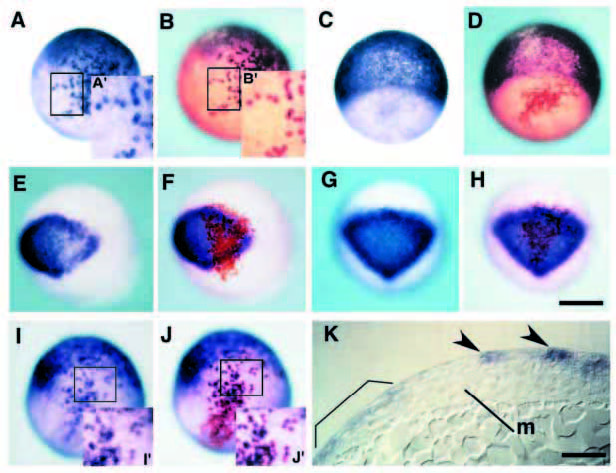
Fig. 7 Constitutively active BMPR-IA can simulate BMP signaling in a direct and cell-autonomous manner in ectoderm patterning. (A-J) Chimeras containing cells derived from injected embryos with biotinylated dextran as a lineage tracer and mRNA encoding either constitutively active receptor (A,B, E,F, I,J) or b-galactosidase (C,D, G,H). Embryos after operation are processed for whole-mount in situ hybridization at 80% epiboly with gata-3, epidermal marker gene (A,C), otx-2, neural marker gene (E,G) and zbmp-2 (I). The same embryos are stained with avidin-biotin HRP complex for the purpose of detection of donor cells (B,D), (F,H), (J), respectively). The insets (A′,B′, I′,J′) show a magnification of the boxed area in the same figures. Note that BMP signaling can activate the expression of gata-3 and zbmp-2 only in cells derived from injected embryo (compare Fig. 7A′ with 7B′ and 7I′ with 7J′, respectively). By contrast, otx-2 is downregulated only in donor cells expressing CABRIA (E,F). In chimeric embryos transplanted cells containing b-galactosidase (C,D, G,H), neither ectopic induction nor repression of marker genes were detected. (K) Sagittal section of chimeric embryos containing CA-BRIA in ectoderm. Ectopic gata-3 signals are observed in ectoderm (arrowheads), suggesting that ectoderm ventralization is independent of mesoderm ventralization. Endogenous gata-3 expression is marked by bracket. Abbreviation: m, axial mesoderm. Scale bar, 200 μm (H), 50 μm (K). A-J are the same magnification.
Image
Figure Caption
Acknowledgments
This image is the copyrighted work of the attributed author or publisher, and
ZFIN has permission only to display this image to its users.
Additional permissions should be obtained from the applicable author or publisher of the image.
Full text @ Development

