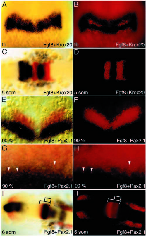
Fig. 3 Fgf8 expression in early midbrain and anterior hindbrain development. (A-D) Double ISH with Fgf8 (blue) and Krox20 (red, fluorescent) of wild-type embryo at tailbud stage (A,B) and 5 somites (C,D). At tailbud stage, Fgf8 expression extends throughout the anterior hindbrain incl. rhombomere 4 (r4) posteriorly. At 5 somites, expression of Fgf8 is detected at the MHB, in r1, r4 and in ventral r2 (see also Fig. 2H). (E-J) Double ISH with Fgf8 (blue) and Pax2.1 (red, fluorescent) at 90% epiboly (E-H) and 6 somites (I,J). At 90% epiboly, the Fgf8 expression domain is located posterior to the Pax2.1 domain with very little overlap (E,F; higher magnification: G,H), while at 6 somites the Fgf8 domain at the MHB is completely included in the Pax2.1 expression domain (visible as quenching of the fluorescent Pax2.1 signal). Embryos in C-J are flat mounted, A,C,E,G and I show bright field, B,D,F,H and J show fluorescent images of the same embryos.
Image
Figure Caption
Figure Data
Acknowledgments
This image is the copyrighted work of the attributed author or publisher, and
ZFIN has permission only to display this image to its users.
Additional permissions should be obtained from the applicable author or publisher of the image.
Full text @ Development

