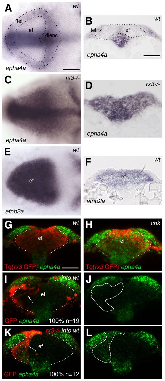
Fig. 1 Complementary expression of Ephrins and Ephs in the anterior neural plate (ANP) is lost in zebrafish rx3-/- mutants. (A-F) Whole-mount in situ hybridisations showing the expression in the ANP of epha4a in wild-type (A,B) and rx3-/- (C,D) embryos and of efnb2a in wild-type embryos (E,F). (G,H) Whole-mount in situ hybridisation to detect epha4a expression in the ANP (green) of Tg{rx3:GFP} (G) and Tg{rx3:GFP}; rx3-/- (chk) (H) embryos counterstained for GFP to highlight the eye field (red). (I-L) Transplants of wild-type (I,J) or rx3-/- (K,L) cells (labelled by GFP, red) into wild-type hosts. rx3-/- cells show autonomous activation of epha4a in the eye field (K,L). Arrows (I,K) point to transplanted cells. Dashed/dotted lines outline ANP domains (A), outline the eye field (B,G-I,K) or outline the transplants (J,L). All panels show frontal views with dorsal to the top of 1- to 3-somite stage (ss) embryos, except (A,C,E) which show dorsal views with anterior to the left. ef, eye field; tel, telencephalon; dienc, diencephalon. Scale bars: 50 μm.
Image
Figure Caption
Figure Data
Acknowledgments
This image is the copyrighted work of the attributed author or publisher, and
ZFIN has permission only to display this image to its users.
Additional permissions should be obtained from the applicable author or publisher of the image.
Full text @ Development

