
Fig. 2
|
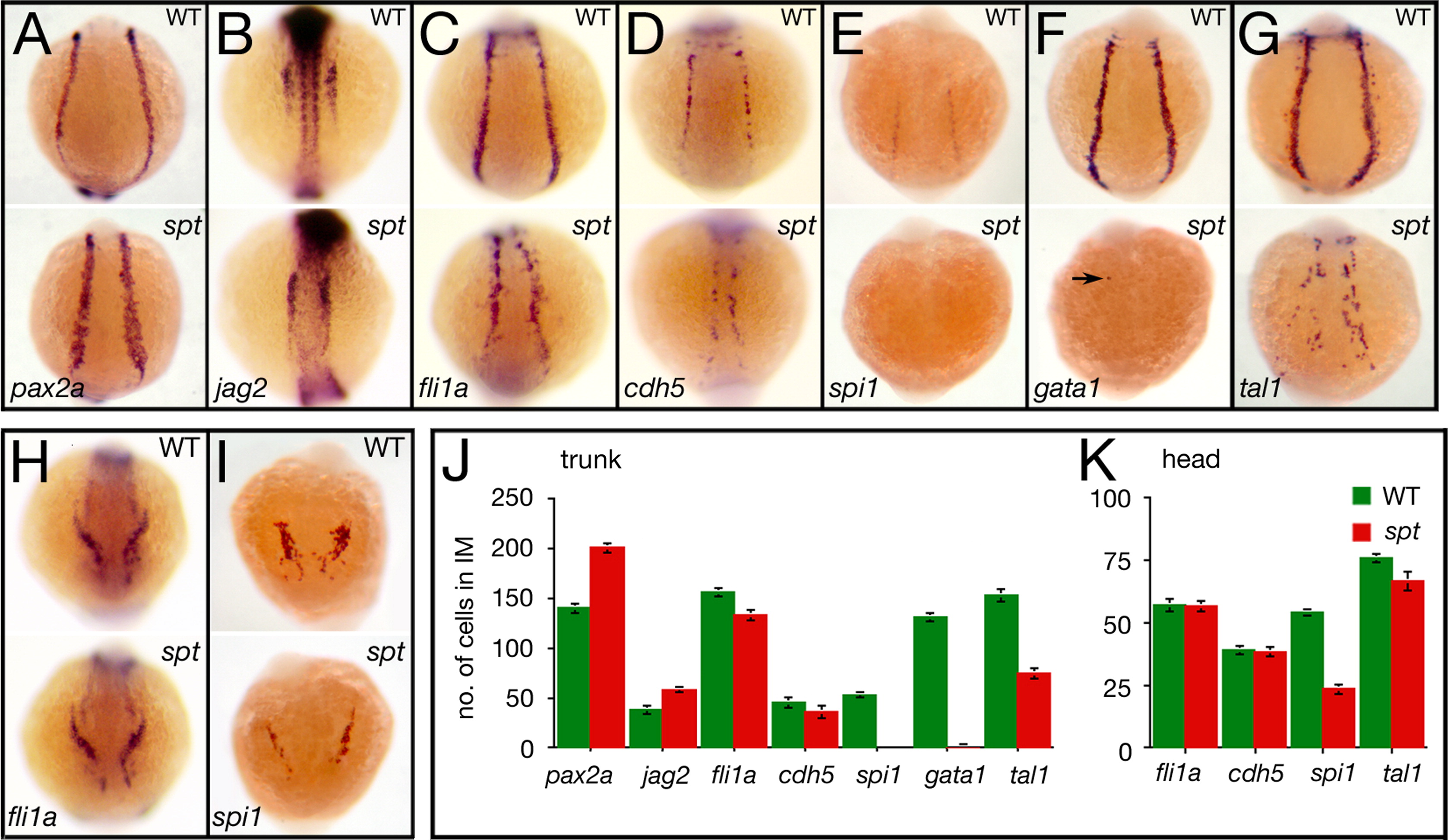
Fig. 2 spt mutants possess more nephric precursors, but have fewer endothelial and hematopoietic precursors. (A?G) Expression in the trunk at 14.5 h of: (A) pax2a and (B) jag2, in nephric cells; (C) fli1a and (D) cdh5, in endothelial cells; and (E) spi1, in white, (F) gata1, in red, and (G) tal1, in all blood cells (arrow designates a single cell). (H, I) Expression in the head at 14.5 h of: (H) fli1a, in endothelial cells; and (I) spi1, in macrophages. (J, K) Quantification of intermediate mesoderm precursors for: (J) trunk, and (K) head. Graphs show the average number of cells per marker including standard error; 9 embryos for each category were counted. Embryos are shown from a (A?G) posterior view, or (H, I) anterior view with dorsal to the top.
Reprinted from Developmental Biology, 383(1), Warga, R.M., Mueller, R.L., Ho, R.K., and Kane, D.A., Zebrafish Tbx16 regulates intermediate mesoderm cell fate by attenuating Fgf activity, 75-89, Copyright (2013) with permission from Elsevier. Full text @ Dev. Biol.