
|
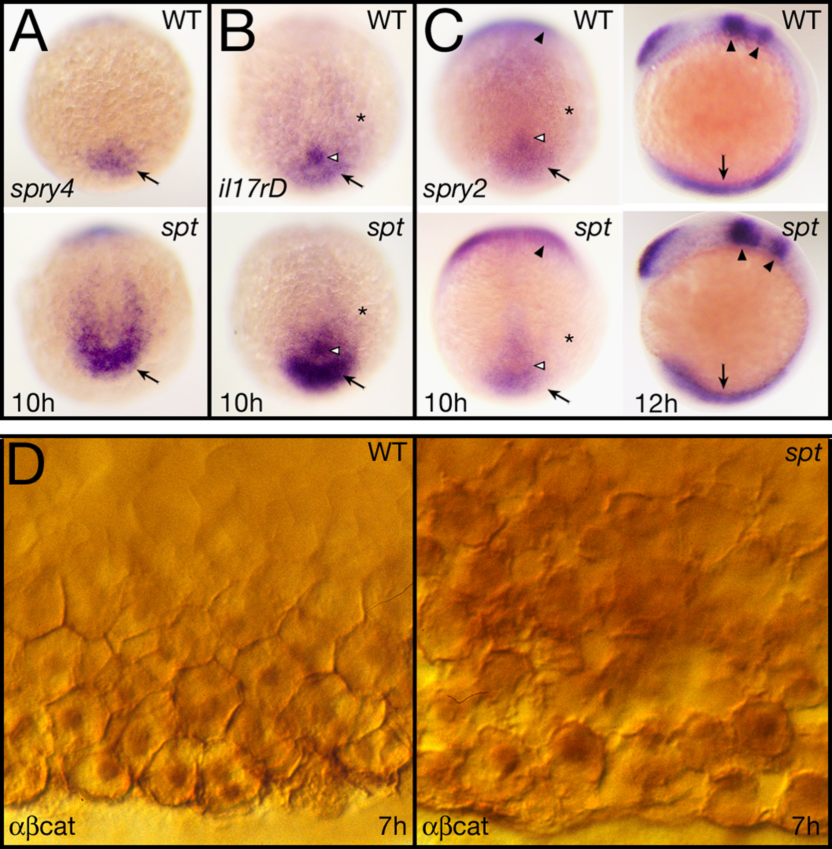
Fig. 7 Fgf and Wnt signaling activity are elevated in spt mutants. (A?C) Expression at 10 and 12 h of: (A) spry4, (B) il17rd, and (C) spry2. All 10 h embryos are shown in a dorsal posterior view, the 12 h embryos in (C) are shown in left side view. Designations: arrow, tailbud (10 h) or posterior mesoderm (12 h); open arrowhead, Kupfer′s vesicle; asterisk, paraxial mesoderm; and arrowheads, midbrain-hindbrain boundary region and rhombomere 4. (D) Expression at 7 h of β-catenin protein seen at high magnification in ventral marginal cells. Cells in the mutant (right), are not as cohesive with one another as in wildtype and make abnormal intercellular contacts and gaps. They also appear to have higher cytoplasmic and nuclear staining.
Reprinted from Developmental Biology, 383(1), Warga, R.M., Mueller, R.L., Ho, R.K., and Kane, D.A., Zebrafish Tbx16 regulates intermediate mesoderm cell fate by attenuating Fgf activity, 75-89, Copyright (2013) with permission from Elsevier. Full text @ Dev. Biol.