
|
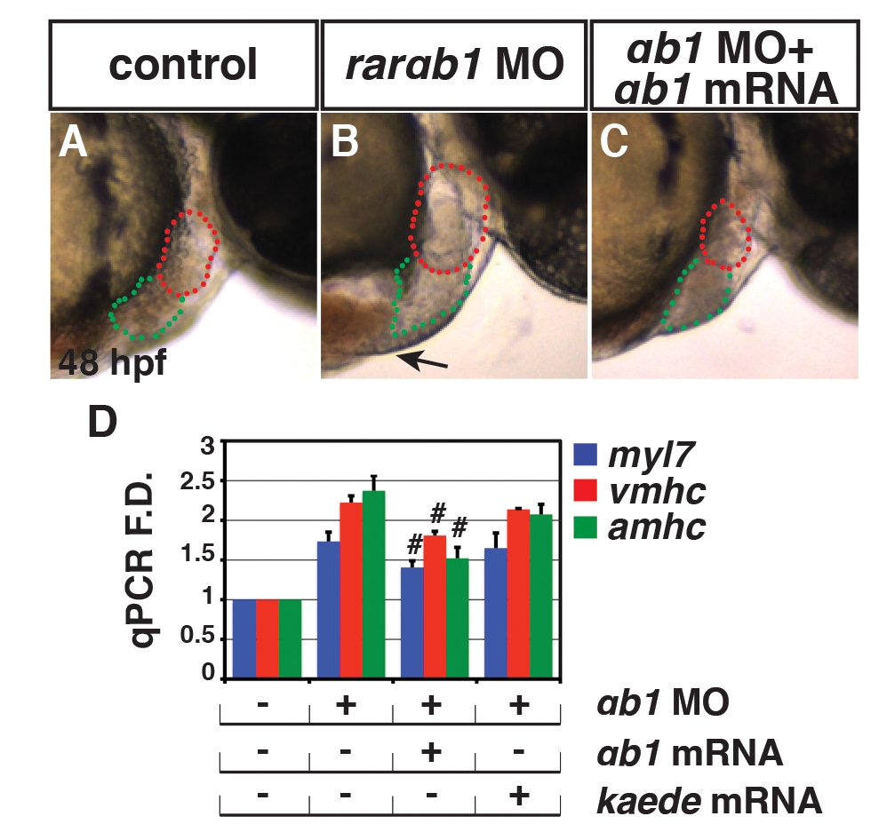
Fig. S4
Specificity controls for the translation blocking rarαb1 MO. (A?C) Control sibling, RARαb1 deficient, and RARαb1 deficient+rarαb1 mRNA injected embryos. Images are lateral views with anterior right at 48 hpf. Red outline indicates ventricles. Green outline indicates atria. Arrow in B indicates edema often found in RARαb1 deficient embryos, which is not found in RARαb1 deficient+rarαb1 mRNA injected embryos (C). (D) qPCR for CM differentiation marker genes at 48 hpf in control sibling, RARαb1 deficient, RARαb1 deficient embryos+rarαb1 mRNA, and RARαb1 deficient embryos+kaede (control) mRNA injected embryos at 48 hpf. Pound sign indicates a statistically significant difference compared to RARαb1 deficient and RARαb1 deficient embryos+kaede (control) mRNA injected embryos (p<0.05).