
|
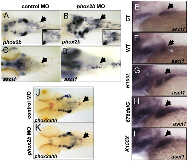
Fig. 5 Deficiency of Phox2b protein due to either MO knockdown or overexpression of PHOX2B variants leads to increased phox2b and ascl1 RNA expression in the SCG.
(A?D) Dorsal views (cranial to the left) of 4-dpf embryos expressing a phox2b ATG MO (B,D) or mismatched control MO (A,C), showing expression of phox2b (A, B) and ascl1 (C, D) as determined by whole-mount ISH. Insets depict an enlarged view of the area of the SCG. (E?I) Area of the SCG is shown in 4-dpf embryos (lateral view, cranial to the left) in which capped mRNA (100 ng/μl) for wild-type (WT) human PHOX2B (F) and the indicated variants (G?I) was injected and ISH performed for ascl1. CT, control, water injected. (J, K) Whole-mount ISH in control vs. phox2b MO-injected embryos double labeled with phox2a (blue) and th (red) riboprobes. Arrows point to the SCG.