
|
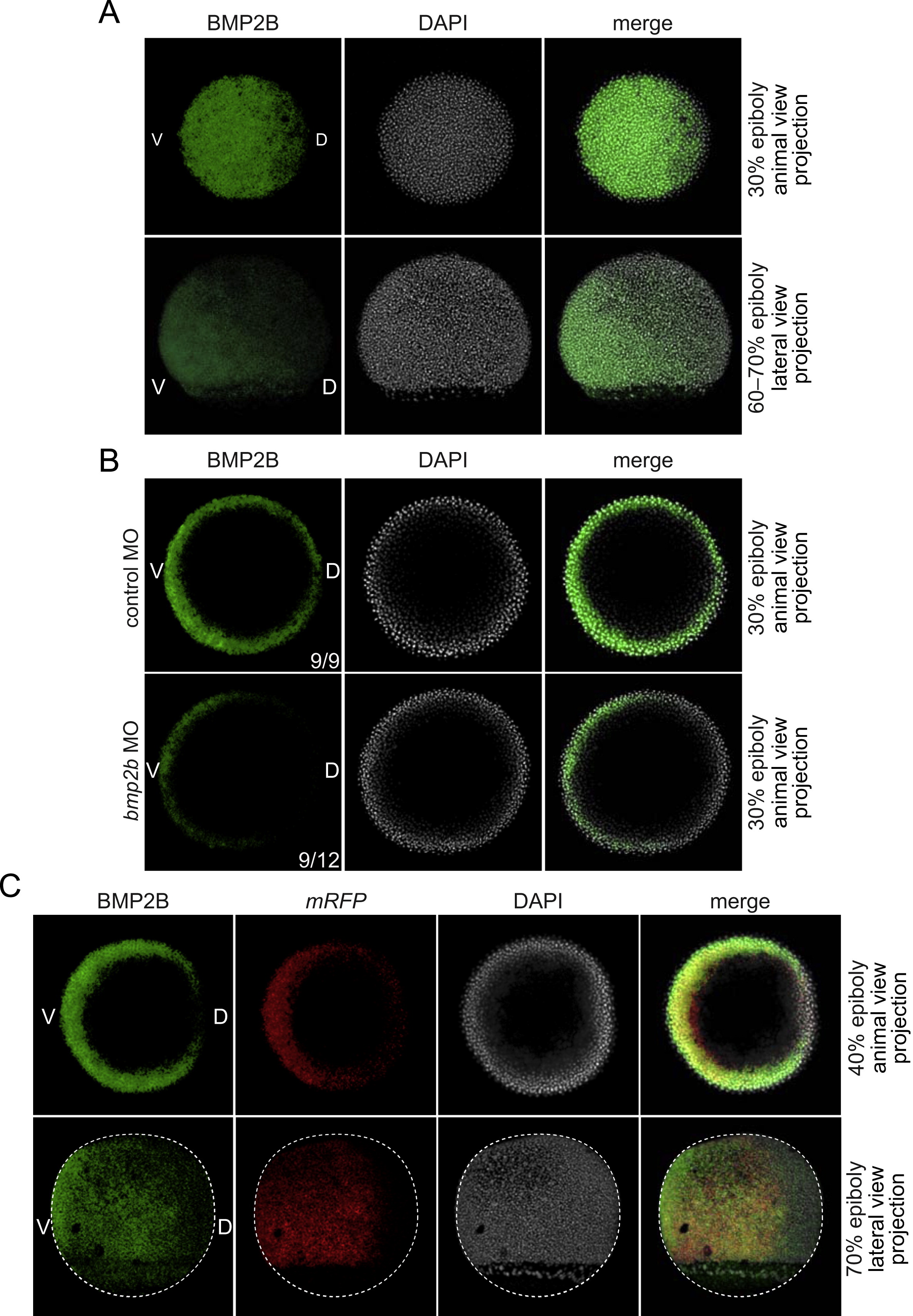
Fig. 5 Graded expression of endogenous BMP2B protein results in graded transcriptional activity. (A) IF for BMP2B in wild-type embryos. (B) IF for BMP2B in control and bmp2b morphants. (C) Combined IF for BMP2B and ISH for mRFP in BRE-mRFP embryos. Note that there is some background staining for BMP2B that is due to autofluorescence from the yolk. Stages and views are indicated. The fluorescent substrate used for mRFP in (C) was Cy5 tyramide (pseudocoloured in red). In (A) and (C, bottom row), the pictures are the projection of a 25?30 stack of images taken from the edge of the embryo until about half way. In (B) and (C, top row), only the blastoderm margin was imaged. The white dotted line in (C, bottom row) shows the outline of the embryo. V, ventral; D, dorsal.
Reprinted from Developmental Biology, 378(2), Ramel, M.C., and Hill, C.S., The ventral to dorsal BMP activity gradient in the early zebrafish embryo is determined by graded expression of BMP ligands, 170-82, Copyright (2013) with permission from Elsevier. Full text @ Dev. Biol.