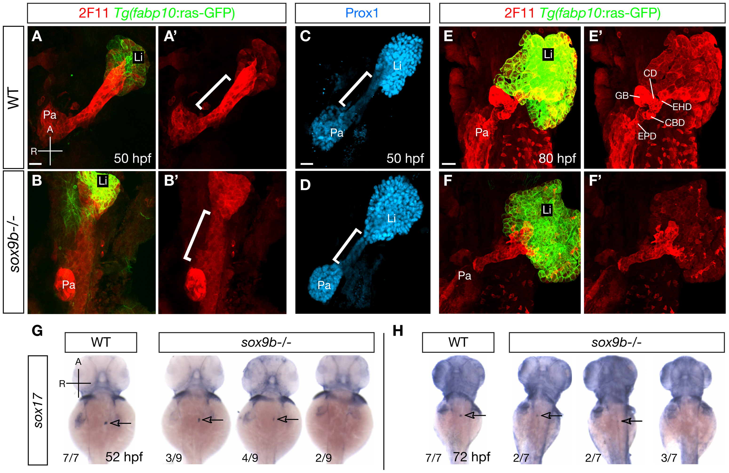
Fig. 3 sox9b mutants display defective HPD patterning during organ development.
(A?B) Labeling for the hepatocyte marker Tg(fabp10:ras-GFP) (green) and the HPD marker 2F11 (red) in wild-type and sox9b mutant embryos at 50 hpf. 2F11 labeling in the HPD primordium is noticeably reduced in sox9b mutants. (A′?B′) Same views as (A?B), but only showing the 2F11 immunostaining. (C?D) In wild-type, Prox1 expression marks the liver and pancreas and is largely absent from the HPD primordium (C). In contrast, Prox1 is abnormally expressed in the HPD primordium in sox9b mutants (D). (A′?B′, C?D) Brackets mark the HPD primordium. (E) By 80 hpf, different compartments of the HPD system, including the cystic duct (CD), common bile duct (CBD), gallbladder (GB), extrahepatic duct (EHD) have become evident in wild-type. (F) In sox9b mutants, the HPD system is dysmorphic and its compartments are indistinguishable based on morphology. The gallbladder is also often missing. (E′?F′) Same views as (E?F), but only showing 2F11 immunostaining. (G?H) Whole-mount in situ hybridization showing sox17 expression in the gallbladder primordium in wild-type and sox9b mutants at 52 (G) and 72 (H) hpf. sox17 expression is greatly reduced or absent in sox9b mutants. The proportion of mutants showing the corresponding phenotype is indicated. Arrows point to the gallbladder primordium. (A?F, A′?F′) All images are projections of confocal z-stacks. Ventral views. (G?H) Dorsal views. Anterior (A) to the top. Pa, pancreas; Li, liver; EPD, extrapancreatic duct. Scale bars, 20 μm.

