
|
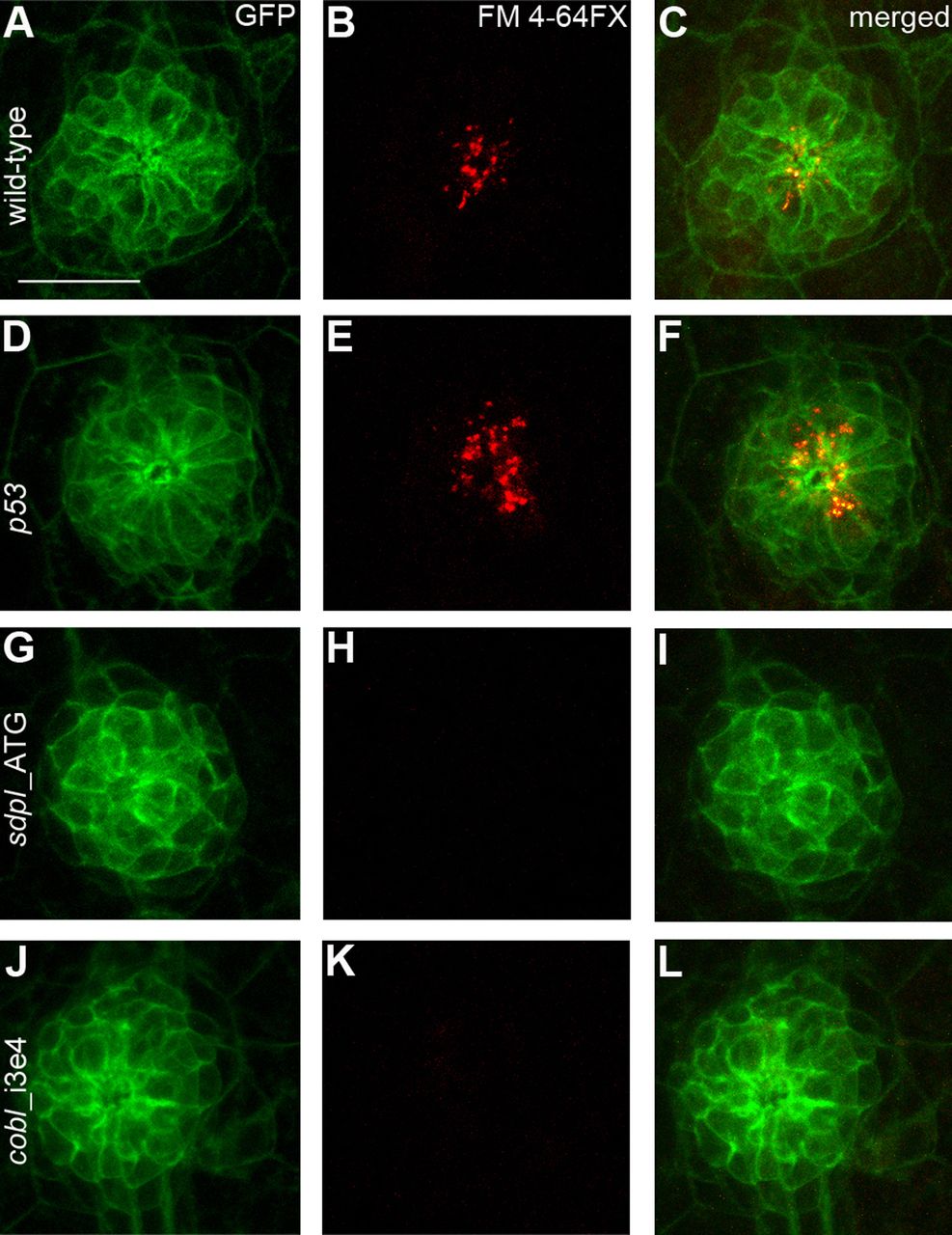
Fig. 5 Sensory hair cells within neuromasts of both cobl- and syndapin I-morphant zebrafish larvae are not functional. (A,D,G,J) Confocal images of representative posterior lateral line neuromasts of wild-type, p53-, sdpI- and cobl-morphant transgenic claudinB:lyn(GFP) larvae at 4dpf. Hair cells are organized symmetrically in rosettes in wild-type (A), control-injected (D), syndapin I (G) and cobl morphants (J). (B,E,H,K) FM 4-64FX vital dye labeling shows a lack of dye uptake in syndapin I (H) and cobl (K) morphants in comparison with wild-type (B) and control-injected larvae (E). (C,F,I,L) Merged images of the mostly apical FM-labeling of vesicular structures and claudin-GFP. Scale bar: 20 μm.