
|
Fig. 6
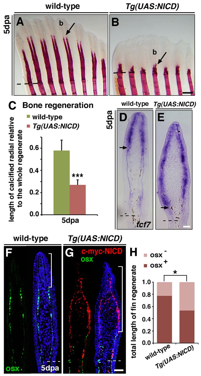
Notch signalling inhibits osteoblast differentiation and prevents bone regeneration. (A,B) Alizarin Red staining of fins at 5 dpa reveals calcified bony rays in a wild-type fin regenerate (A, arrow). Fin rays are poorly formed in Tg(UAS:NICD) fins, whereas the blastema (b) is expanded (B, arrow). (C) Length of regenerated calcified radials of Tg(UAS:NICD) fins, relative to wild-type fins. ***P<0.001. (D,E) In situ hybridization at 5 dpa. Wild-type fins express tcf7 in peripheral cells in the distal region (D, arrow), whereas transcription is expanded proximally in Tg(UAS:NICD) fins (E, arrow). (F,G) Immunostaining for Osx and Myc-NICD. In wild-type fins, Osx+ cells (green) are present in the proximal fin region but scarce in the distal region (F, bracket). Osx+ cells are less numerous in regenerating Tg(UAS:NICD) fins and more proximally limited. The region of high Myc-NICD expression does not contain Osx+ cells (G, bracket). (H) Relative length of the proximal region of the fin regenerate, which contains Osx+ cells, and the distal region (brackets, F,G), which does not (Osx-). The distal, Osx- region is larger in Tg(UAS:NICD) fins than in wild-type fins. *P<0.05. Scale bars: 200 μm in B; 10 μm in E,G. Broken lines indicate the amputation plane.