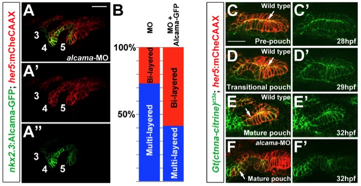
Fig. S4 Requirement of Alcama in endoderm and its role in α-catenin localization, related to Figure 5.
(A) Transgenic expression of Alcama-GFP in nkx2.3:Alcama-GFP; her5:mCherryCAAX embryos partially restores bilayered pouch morphology to alcama-MO-injected embryos. Alcama-GFP (green) localizes to apical and lateral membranes of pouches labeled with her5:mCherryCAAX (red). Pouches 3-5 are numbered. Scale bar = 20 7mu;M. (B) Quantification of rescue of alcama-MO pouch defects by Alcama-GFP transgenic expression. Pouches 4 and 5 were used for the analysis. n=119 for alcama-MO + Alcama- GFP, and n=118 for alcama-MO alone. p-value = 0.000001. (C-E) Still images from a time-lapse confocal recording of fifth pouch development in a Gt(ctnna-citrine)ct3a; her5:mCherryCAAX embryo (See Movie S7). In the pre-pouch endoderm, &alpjha;-catenin-citrine (green) is enriched along apical membranes (arrows) of the her5:mCherryCAAX-labeled endodermal bilayer (red). During pouch initiation, α-catenincitrine becomes less organized in the multi-layered transitional epithelium. As pouches mature, α-catenin-citrine becomes organized again along apical membranes in the bilayer. (F) At a stage when wild-type pouches would have matured into bilayers, α-catenin-citrine remains disorganized in the alcama-MO multilayered pouches (arrow).
Reprinted from Developmental Cell, 24(3), Choe, C.P., Collazo, A., Trinh, L.A., Pan, L., Moens, C.B., and Crump, J.G., Wnt-Dependent Epithelial Transitions Drive Pharyngeal Pouch Formation, 296-309, Copyright (2013) with permission from Elsevier. Full text @ Dev. Cell

