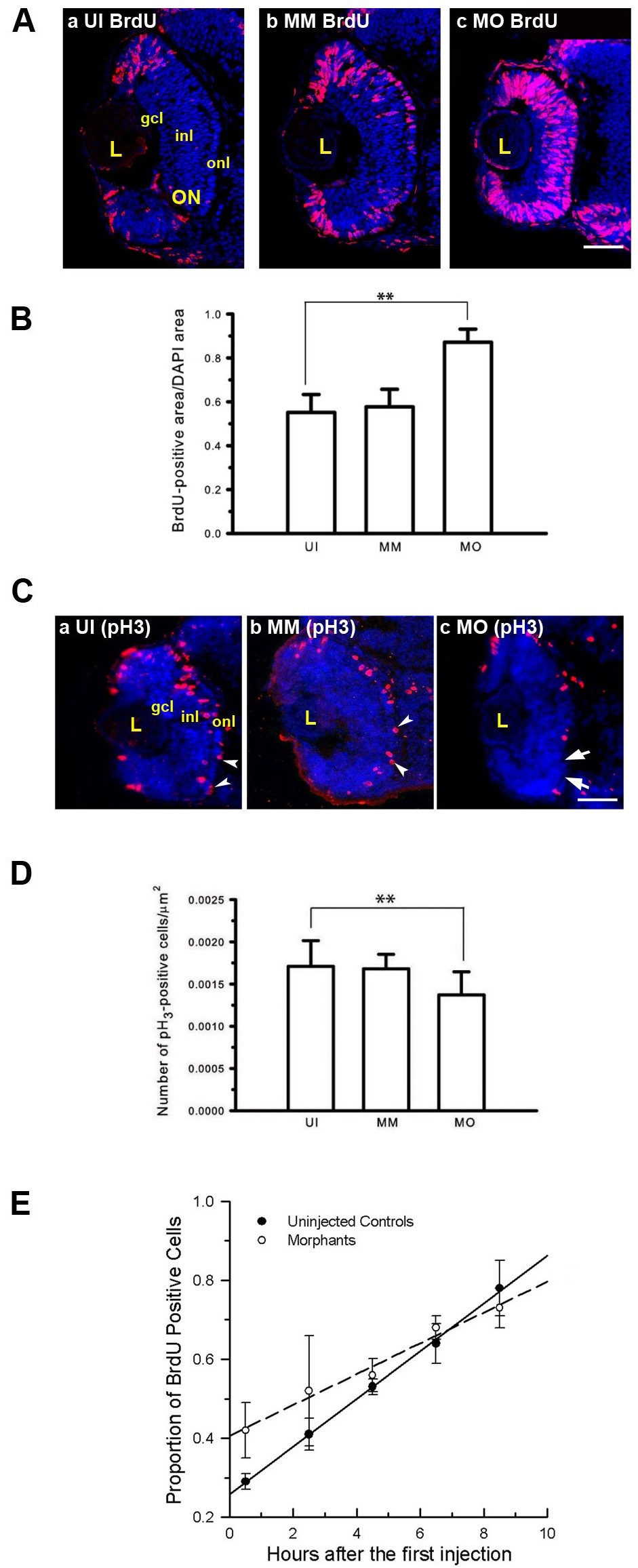
Fig. 3 Mdka loss-of-function alters cell cycle kinetics. (A) BrdU labeled progenitors in uninjected (a) control (b) and Mdka loss-of-function embryos (c). (B) Histograms showing the average proportion of the retinal section occupied by BrdU-labeled cells. *P <0.001. n = 9 retinas/group. Scale bar equals 50μm.(C) Retinas of embryos at 48 hpf stained with antibodies against phosphohistone H3 (pH3) in uninjected (a), control (b), and Mdka loss-of-function embryos (c). (D) The number of pH3-stained cells/unit area. **P <0.01. n=12 embryos/group. MM, embryos injected with 5-pair mismatch control morpholinos; MO, embryos injected with mdka-targeted morpholinos (MO); UI, uninjected embryos. gcl, ganglion cell layer; inl, inner nuclear layer; L, lens; ON, optic nerve; onl, outer nuclear layer. Scale bar equals 50 μm. (E) Graph of the least-squares regression lines through data showing the proportion of BrdU-labeled progenitors as a function of time. An F-test was used to compare the control and experimental data. **P <0.05.
Image
Figure Caption
Figure Data
Acknowledgments
This image is the copyrighted work of the attributed author or publisher, and
ZFIN has permission only to display this image to its users.
Additional permissions should be obtained from the applicable author or publisher of the image.
Full text @ Neural Dev.

