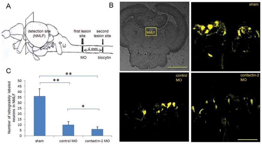
Fig. 6 Reduction of contactin-2 protein expression after spinal cord injury impairs axon regrowth of NMLF neurons.
(A) Schematic illustration for retrograde tracing. Biocytin was applied at the site symbolized by the second lesion site at 6 weeks after the first lesion, and was detected in the NMLF after 24 hrs using Streptavidin-Cy3. (B) Retrogradely labeled neurons in the NMLF. (C) Numbers of retrogradely labeled neurons in the NMLF. Reduction of contactin-2 protein expression by application of contactin-2 anti-sense MO reduces the numbers of NMLF neurons with axons regrown beyond the first lesion site when compared to the control MO treated group (*p<0.05, t-test; n = 3 fish/group). The sham injury group shows similar numbers of retrogradely labeled neurons as the non-injury group. Scale bars, 200 μm and 50 μm.

