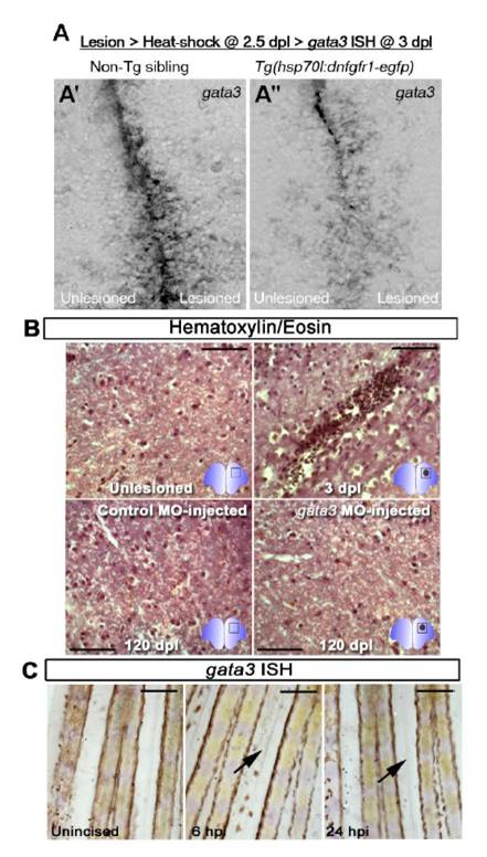
Fig. S4 (A-A′ ′) Transient and brief block of Fgf signalling reduces gata3 expression significantly. (A) Heat shock and lesion scheme. (A′) gata3 in situ hybridization on adult telencephalic ventricle of the heat-shocked non-transgenic siblings. gata3 expression is strong and seen in ventricular cells. (A′ ′) gata3 in situ hybridization on adult telencephalic ventricle of the heat-shocked Tg(hsp:dnfgfr1-egfp) animals. gata3 expression reduces significantly in the ventricular cells. (B) Hematoxylin and Eosin staining on unlesioned and 3 dpl adult zebrafish brain indicates blood clotting and and tissue remodelling after lesion. At 120 dpl, control morpholino-injected brains display a tissue architecture reminiscent of the unlesioned brain. Gata3 morphant brains also show restored tissue architecture, indicating that Gata3 function at the ventricular region does not affect the gliotic response, which normally resolves quickly to enable regenerative neurogenesis. (C) In intact (unincised) adult zebrafish caudal fin, gata3 is not detectable. gata3 expression is not induced after incision and during wound healing at 6 and 12 hours after incision (hpi). n = 4 fish for A-A′ ′, 6 for every panel in B, 3 for every panel in C.
Reprinted from Developmental Cell, 23(6), Kizil, C., Kyritsis, N., Dudczig, S., Kroehne, V., Freudenreich, D., Kaslin, J., and Brand, M., Regenerative Neurogenesis from Neural Progenitor Cells Requires Injury-Induced Expression of Gata3, 1230-1237, Copyright (2012) with permission from Elsevier. Full text @ Dev. Cell

