
|
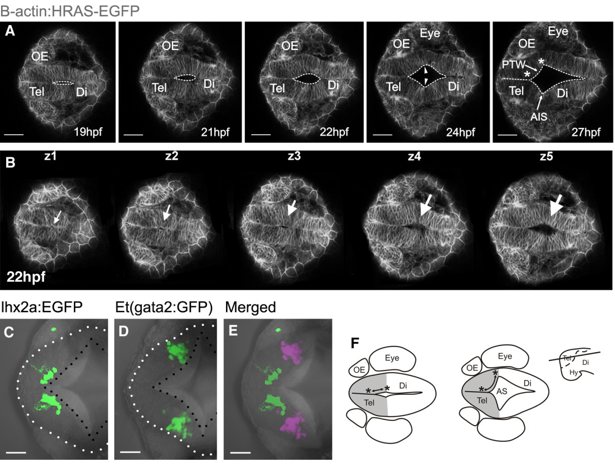
Fig. 2 Formation of anterior intraencephalic sulcus (AIS) and posterior wall of the telencephalon. A. Time-lapse sequence of a single z-level of a Tg(β-actin: HRAS-EGFP) vu119 embryo. The midline lumen opens out at the telencephalic-diencephalic boundary to form the AIS and the posterior wall of the telencephalon (ventricular surface between asterisks). Inflation of the ventricular space leads to a lateral protrusion of the telencephalon in its caudal region (arrowheads). B. Five z-levels at a single timepoint showing opening of the AIS (arrowed) is initiated deep (z5) from the dorsal surface (z1). C. Cells of the olfactory bulb, revealed in Tg(10lhx2a:EGFP)zf176 line, lie medially and just in front of the posterior telencephalic wall at 30 hpf. D. Pallial cells in the Et(gata2:GFP)bi105 line lie laterally, just in front of the posterior telencephalic wall at 30 hpf. E. Olfactory bulb [Tg(10lhx2a:EGFP)zf176] and pallial cells [Et(gata2:GFP)bi105] in C and D superimposed to show relative positions (pallial cells pseudocoloured turquoise). F. Diagram to illustrate how generation of AIS folds posterior telencephalon to a more lateral location. Dorsal views, anterior to the left. Scale bars: 100 μm in A and B and 50 μm in B to E. AIS: anterior intraencephalic sulcus; Di: diencephalon; Hy: hypothalamus; OE: olfactory epithelium; PTW: posterior telencephalic wall; Tel: telencephalon.