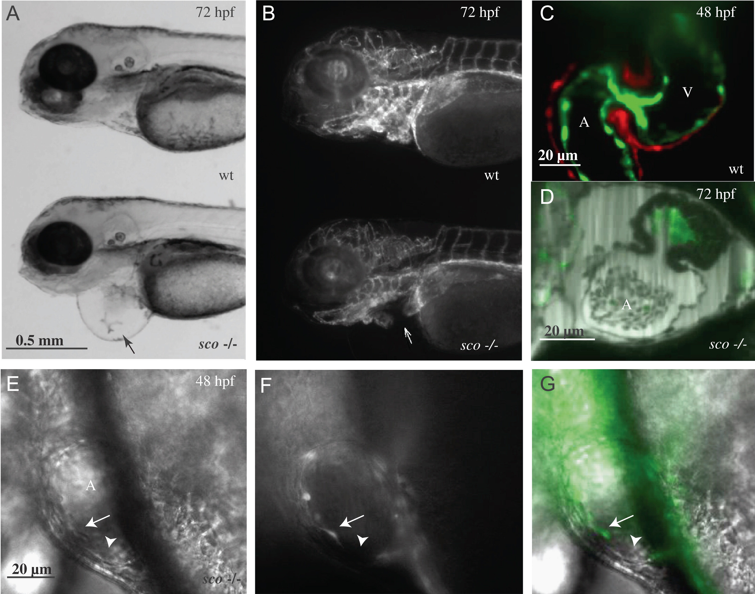
Fig. 2 Heart vasculature defects in scote382 mutants. Fluorescence images of 72 hpf Tg(kdrl:GFP)s843 wild-type (A) and scote382 mutant (B) larvae. The overall patterning of the vasculature appears unaffected in scote382 mutant larvae; however, pronounced pericardial edema (black arrow in panel A) indicates a severe defect in heart function. Closer inspection reveals the absence of endocardial cells in the mutant atrium (white arrow in panel B). SPIM images show Tg(kdrl:GFP)s843; Tg(my17:DsRed)s879 wild-type endocardium and myocardium (C) at 48 hpf and the Tg(kdrl:GFP)s843scote382 mutant atrium, which appears to lack its endocardial lining (D) at 72 hpf. Panels C and D are stills taken from Video 1 and Video 2, respectively. Panels E?G are DIC images of a scote382 mutant atrium taken at 40×. An endocardial cell (white arrow) fails to adhere to its neighbor, thereby creating a gap (white arrowhead) in the endocardial sheet; DIC (E), the endothelial kdrl:GFP reporter (F) and merged image (G). A: atrium; V: ventricle.
Reprinted from Developmental Biology, 372(1), Mellman, K., Huisken, J., Dinsmore, C., Hoppe, C., and Stainier, D.Y., Fibrillin-2b regulates endocardial morphogenesis in zebrafish, 111-119, Copyright (2012) with permission from Elsevier. Full text @ Dev. Biol.

