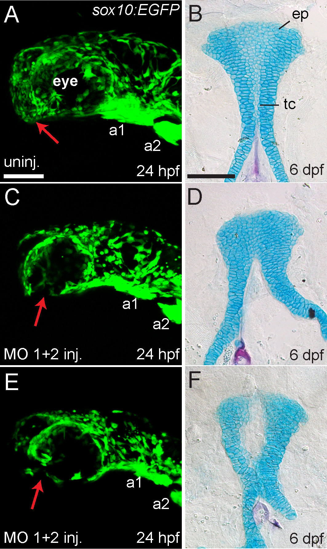
Fig. 6
Reduction or absence of neural crest cells in MO-injected embryos is associated with palatal defects. (A,C, E) Lateral views of live sox10:EGFP transgenic embryos at 24 hpf. Anterior is towards the left, dorsal is upwards. (B,D,F) Ventral view of Alcian Blue (cartilage) and Alizarin Red (bone) stained palatal skeletons of the same individual fish shown in A,C,E, fixed and flat-mounted at 6 dpf. Anterior is upwards. (A) Uninjected embryos (n = 7) showed GFP expression in neural crest-derived tissues in the head including arch 1 (a1) and arch 2 (a2), and GFP-positive cells populate the anterior-ventral margin of the face where the palatal skeleton forms (indicated by red arrow). (B) Uninjected embryos had normal development of the ethmoid plate (ep) and trabeculae communis (tc) of the palatal skeletons. (C and E) hdac4 MO-injected embryos (n = 6) showed GFP expression in neural crest-derived tissues in the head, including arch 1 (a1) and arch 2 (a2), but a lack of cells in the anterior-ventral margin of the face at 24 hpf (indicated by red arrows) (D and F) The same hdac4 MO-injected embryos in C and E had palatal skeleton defects at 6 dpf (n = 6/6), including defects in formation of the trabeculae communis (D), and ethmoid plate (F). A,C,E scale bars = 100 μm, B,D,F scale bar = 100 μm.

