
Image
|
Figure Caption
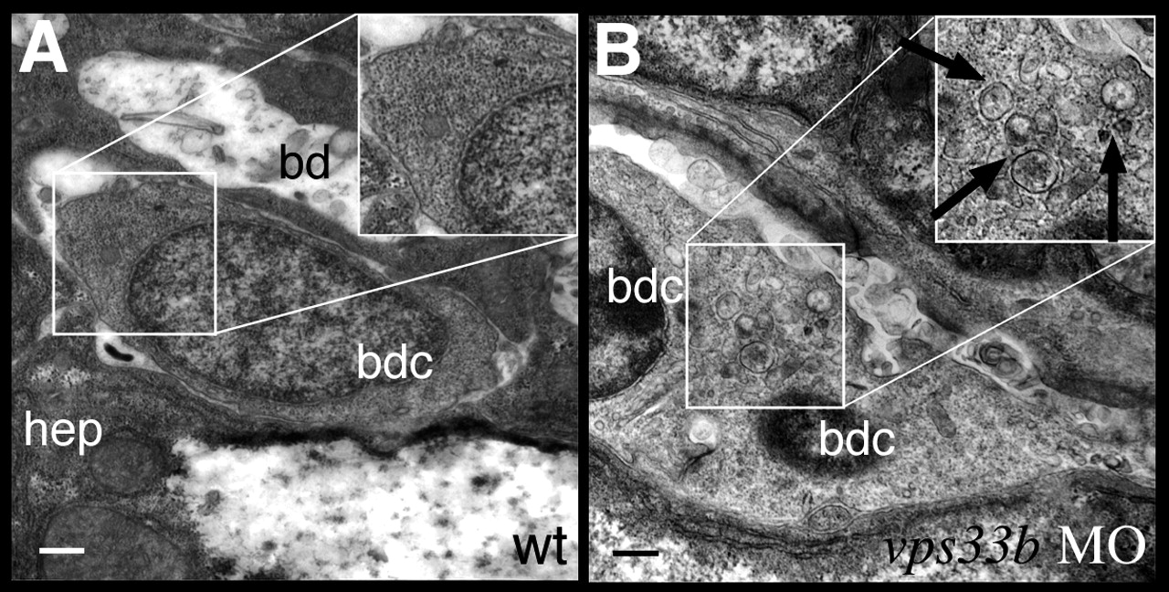
Fig. 5
vps33b knockdown disrupts biliary ultrastructure. (A,B) Electron micrographs of biliary epithelial cells from 5 dpf wild-type (A) and vps33b morpholino injected (B) larvae. The wild-type biliary cell cytoplasm has a homogeneous appearance. (B) A small bile duct comprising two bile duct cells from a vps33b morpholino-injected larva. Cytoplasm appears heterogeneously with multiple vesicles (black arrows). hep, hepatoctye; bd, bile duct lumen; bdc, bile duct cell.
Figure Data
Acknowledgments
This image is the copyrighted work of the attributed author or publisher, and
ZFIN has permission only to display this image to its users.
Additional permissions should be obtained from the applicable author or publisher of the image.
Full text @ Development