Fig. 4
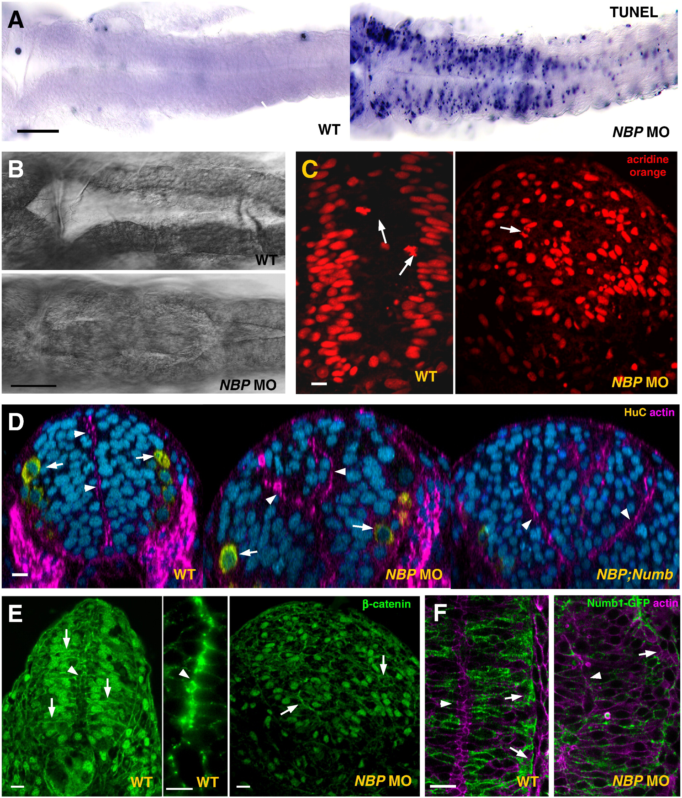
NBP is required for neural tube development. Panels compare NBP morphants (NBP MO) and double knockdown NBP;Numb mutants (NBP;Numb) with wild-type embryos (WT). (A) Many cells in the neural tube of the 24 hpf morphants, particularly of the embryos injected with 1 mM NBP MO, gave positive reaction in the TUNEL assay when compared to embryos either injected with the mismatch NBP MOC (at 1 mM, not shown) or un-injected (flat preparations, anterior to the left). (B) Abnormal growth in the ventricle of the hindbrain in NBP MO embryos (flat preparations, anterior to the left). (C?F) Neural tube defects in the NBP morphants. In embryos depleted from NBP (E, right picture) the cells of the neural tube frequently failed to form the usual epithelial structure composed of elongated columnar cells (arrows point to β-catenin membrane staining of baso-apically oriented polarized wild-type cells and of randomly oriented rounded cells in the morphant), the tissue appeared to be unorganized and did not form the typical midline enriched in β-catenin and actin ring characteristic for wild-type (D; E, arrowheads in the left and middle pictures). The nuclei (visualized by acridine orange) were not placed in their characteristic positions (C, left picture) but were scattered in the whole volume and their divisions were not restricted to the apical side of the neural tube (arrow in C, right picture, points to an anaphase in a deep region of the neural tube). For the morphant, E and C represent the same section probed by anti-β-catenin antibody (E) and acridine orange (C), respectively. (D) More than one neurocoel developed in the NBP morphants (middle panel), a phenotypic aberration enhanced in the NBP;Numb double morphants (arrowheads, neurocoel; arrows; HuC positive cells). (F) In the NBP morphant, Numb1 did not display the typical accumulation at the basal pole of cells (arrowhead, midline; arrow, basal pole of the neural cells). Scale bar: A, B = 100 μm, and C?F = 5 μm.
Reprinted from Developmental Biology, 365(1), Boggetti, B., Jasik, J., Takamiya, M., Strähle, U., Reugels, A.M., and Campos-Ortega, J.A., NBP, a zebrafish homolog of human Kank3, is a novel Numb interactor essential for epidermal integrity and neurulation, 164-174, Copyright (2012) with permission from Elsevier. Full text @ Dev. Biol.

