
Fig. 5
|
Fig. 5
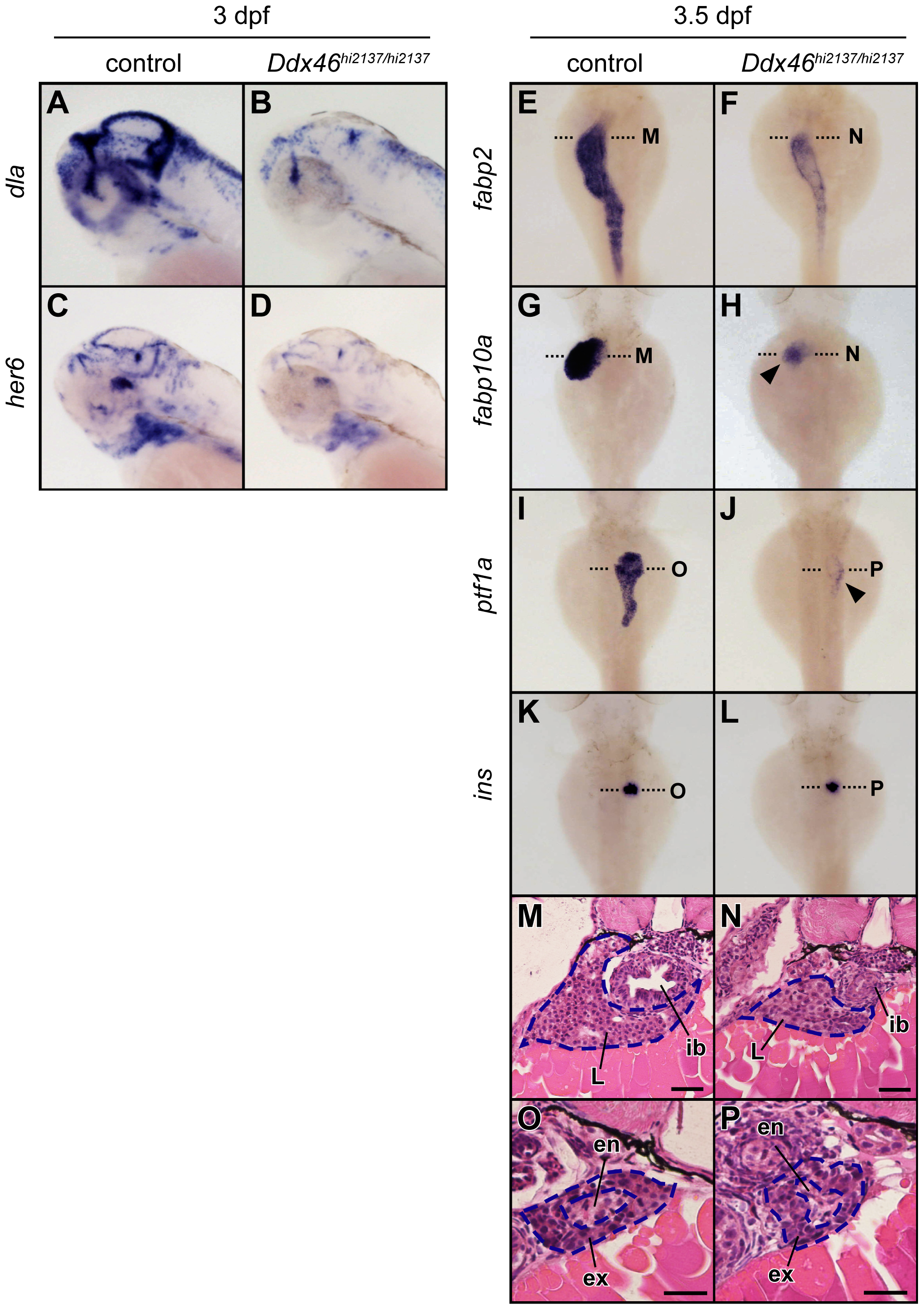
Expression of molecular markers for digestive organs and brain is reduced in the Ddx46hi2137/hi2137 mutant.
(A?D) The expression of dla and her6 was examined using whole-mount in situ hybridization at 3 dpf. All lateral views, anterior to the left. (E?L) The expression of fabp2, fabp10a, ptf1a, and ins was examined using whole-mount in situ hybridization at 3.5 dpf. All dorsal views, anterior to the top. In the Ddx46hi2137/hi2137 mutants, the intensity and area of dla, her6, fabp2, fabp10a, and ptf1a expression were markedly reduced at 3 or 3.5 dpf (A?J; arrowheads in H, J). In contrast, the ins expression in the Ddx46hi2137/hi2137 mutant did not change at these developmental stages (K, L). (M?P) Transverse sections of 3.5-dpf Ddx46hi2137/hi2137 mutant larvae stained with hematoxylin and eosin. The transverse sections were cut at the levels indicated by black dotted lines in E?L. The tissues in the intestinal bulb, liver, and exocrine pancreas were still present in the Ddx46hi2137/hi2137 mutant larvae at 3.5 dpf. Scale bars, 50 μm. en, endocrine pancreas; ex, exocrine pancreas; ib, intestinal bulb; L, liver. Control larvae were sibling WT or Ddx46hi2137/+ larvae and had normal phenotypes.