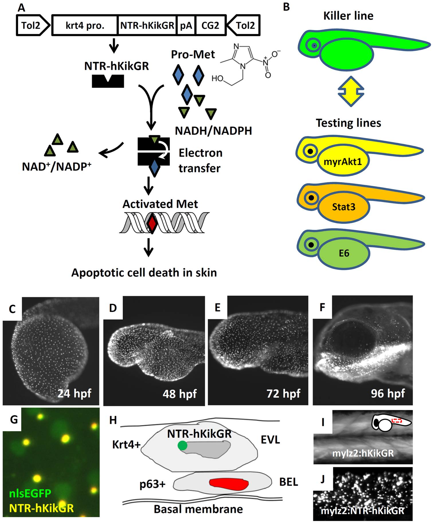
Fig. 2 Establishment of Tg(krt4:NTR-hKikGR)cy17 killer line.
(A) The work flow to conditionally ablate zebrafish skin using NTR/Met-mediated system. The superficial skin-specific krt4 promoter controls NTR-hKikGR fusion protein. Tol2 transposon elements flank the whole transgene cassette and enhance the germ-line transmission rate. Dimerization of NTR-hKikGR transfers electrons from NADH/NADPH to Met prodrug. Activated Met crosslinks DNA and specifically triggers apoptotic death in skin. (B) The killer line carrying the krt4:NTR-hKikGR transgene was crossed with several testing lines which overexpress human constitutively active myrAkt1 (myrAkt1), mouse constitutively active Stat3 (Stat3), or HPV16 E6 (E6) genes. The double transgenics were then subjected to Met incubation to assay the potential function of apoptosis modulators. (C?F) The ontogenic expression pattern of NTR-hKikGR fusion protein in killer line aged from 24 to 96 hpf. (G) The living fluorescent signals detected in double transgenics from the crossing of the killer line and Tg(krt4:nlsEGFP)cy34 aged at 72 hpf show that the NTR-hKikGR+ signals (yellow) aggregated adjacently to the nlsEGFP+ (green) skin nucleus. (H) Model to illustrate the spatial distribution of NTR-hKikGR fusion protein in zebrafish embryo skin. (I) The native hKikGR protein displays cytoplasmic distribution pattern in the skeletal muscle of Tg(mylz2:hKikGR). The relative position of the captured image is highlighted at the upper right corner. (J) The NTR-hKikGR fusion protein aggregated in skeletal muscle of Tg(mylz2:NTR-hKikGR). EVL, enveloping layer; BEL, basal epidermal layer; Met, metrodinazole.

