Fig. 5
Fig. 5
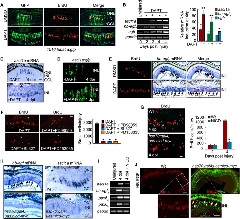
Notch Inhibition Stimulates MG Proliferation and Expands the Zone of Dedifferentiated MG in the Injured Retina via Induction of hb-egfa Gene Expression (A) DAPT treatment of 1016 tuba1a:gfp transgenic fish results in an expansion of the zone of dedifferentiated MG (GFP+/BrdU+) in the injured retina. Scale bar, 50 μm. (B) RT-PCR (gel) and real-time PCR (4 dpi, graph) showing that DAPT induces ascl1a, hb-egfa, and egfr mRNA expression in the injured retina. **p < 0.01 for ascl1a and hb-egfa; *p < 0.05 for egfr. (C) In situ hybridization showing that DAPT expands ascl1a mRNA expression in the injured retina (* marks the injury site). Scale bar, 50 μm. (D) ascl1a:gfp transgenic fish exhibit injury- and Notch-dependent regulation of transgene expression. Scale bar, 50 μm. (E) DAPT stimulates hb-egfa mRNA expression in proliferating MG-derived progenitors (arrows) of the injured retina. Scale bar, 50 μm. (F) MAPK inhibition (PD98059 and SL327) and EGFR inhibition (PD153035) suppress DAPT-dependent expansion of MG proliferation in the injured retina (asterisks in photomicrographs identify injury sites). Graph quantifies data represented in photomicrographs. *p < 0.01. (G) BrdU immunofluorescence on retinal sections reveals BrdU incorporation in the injured retinas of WT and hsp70:gal4;uas:nicd-myc transgenic fish overexpressing NICD. Scale bar, 50 μm. *p < 0.01. (H) In situ hybridization assays for hb-egfa (arrows) and ascl1a mRNA expression in WT and hsp70:gal4;uas:nicd-myc fish overexpressing NICD (* marks the injury site). Scale bar, 50 μm. (I) RT-PCR shows that ascl1a, hb-egfa, and pax6b mRNA expression is inhibited by NICD overexpression. (J) NICD-myc overexpression suppresses HB-EGF-induced cell proliferation in the uninjured retina. BrdU+ cells in HB-EGF-treated retinas overexpressing NICD-myc are generally NICD negative (arrows). Scale bars are 150 μm in low-magnification images and 50 μm in higher-magnification images. Error bars represent SD. ONL, outer nuclear layer; INL, inner nuclear layer; GCL, ganglion cell layer. See also Figure S6.
Reprinted from Developmental Cell, 22(2), Wan, J., Ramachandran, R., and Goldman, D., HB-EGF Is Necessary and Sufficient for Müller Glia Dedifferentiation and Retina Regeneration, 334-347, Copyright (2012) with permission from Elsevier. Full text @ Dev. Cell

