
Fig. 1
- ID
- ZDB-IMAGE-111201-25
- Genes
- Antibodies
- Source
- Figures for Bachmann-Gagescu et al., 2011
|
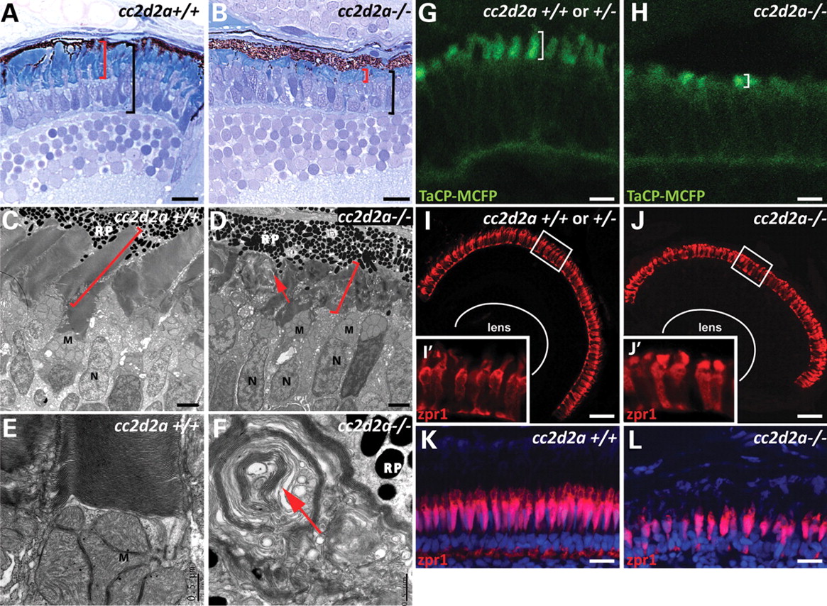
Fig. 1 cc2d2a is required for zebrafish photoreceptor outer segment development. (A and B) Plastic sections of 5 d.p.f. cc2d2a+/+ (A) and cc2d2a-/- (B) retinas. Black brackets highlight the photoreceptor cell layer, and red brackets highlight the outer segments. (C?F) Transmission electron microscopy on 5 d.p.f. retinal sections shows disorganized stacks of membrane (red arrow) in the outer segments of cc2d2a-/- retinas (D) compared with wild-type (C). Higher power views (E and F) show whorls of membrane stacks replacing the outer segment (red arrow). (G and H) Tg(TaCP:MCFP) expression in 80 h.p.f. cryosectioned retinas. Wild-type cc2d2a+/+ or +/- cone photoreceptors have readily visible outer segments (G), whereas cc2d2a-/- photoreceptors have only short outer segments without the typical shape at the same stage (H). (I?J′) Red?green cones labeled with zpr1 antibody in wild-type (I and I′) and cc2d2a-/- (J and J′) 5 d.p.f. retinal cryosections showing comparable density of cone photoreceptors and grossly preserved photoreceptor cell morphology except in the apical segment. (K and L) Red?green cone cell bodies labeled with zpr1 antibody (red) in retinal cryosections from 4-week-old wild-type (K) and cc2d2a-/- fish (L). Fluorescent images are single-confocal sections. M, mitochondria; N, nuclei; RP, retinal pigment. Scale bars are 10 �m in (A and B), 2 �m in (C and D), 0.5 �m in (E and F), 4 �m in (G and H), 20 �m in (I and J) and 10 �m in (K and L).