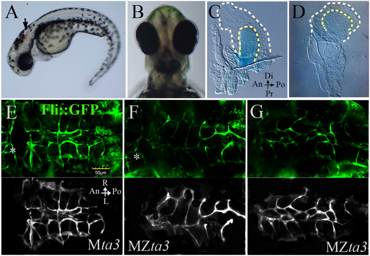
Fig. 5 Fin and head abnormalities in MZta3 mutants. (A,B) MZta3 embryos at 2 dpf displaying hindbrain haemorrhage (arrow) and mild cyclopia (B). (C,D) Pectoral fins from Mta3 and MZta3 larvae stained with Alcian Blue, showing loss of asymmetry in mutant (D). Broken lines indicate the fin fold margin. (E-G) Cranial blood vessels marked by the fli::GFP transgene; background had been removed in lower monochrome panels for ease of observation. Note the mis-branched and merged vessels of the cerebellar central artery in the MZta3 embryos (F,G), beneath the haemorrhagic area. Posterior mesencephalic central artery is indicated by an asterisk. An, anterior; Po, posterior; Di, distal; Pr, proximal; L, left; R, right.
Image
Figure Caption
Figure Data
Acknowledgments
This image is the copyrighted work of the attributed author or publisher, and
ZFIN has permission only to display this image to its users.
Additional permissions should be obtained from the applicable author or publisher of the image.
Full text @ Development

