Fig. 7
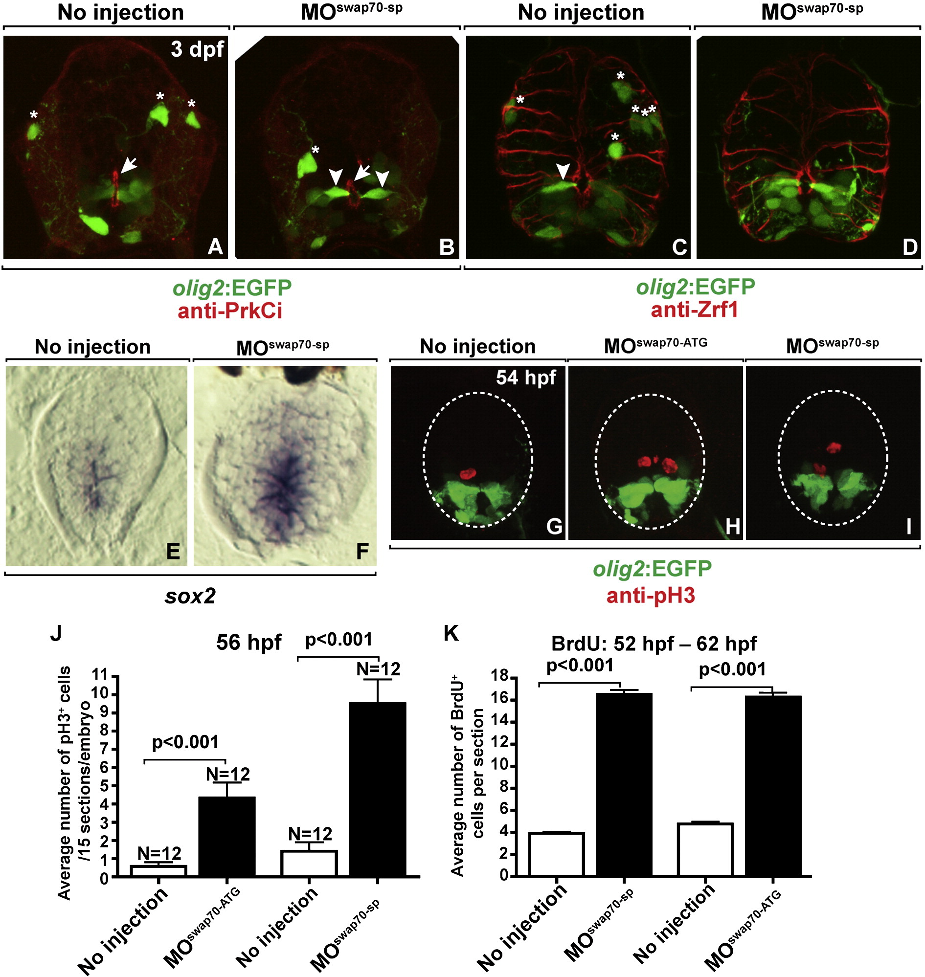
swap70 loss-of-function does not dramatically alter spinal cord development. All images show transverse sections through trunk spinal cord with dorsal up. (A, B) Anti-PrkCi immunocytochemistry performed on control and MOswap70-sp-injected larva at 3 dpf. PrkCi labeling of apical membrane (arrows) and EGFP labeling of olig2+ radial glia (arrowheads) is indistinguishable between control and experimental animals whereas fewer EGFP+ OPCs (asterisks) are evident following MOswap70-sp injection. (C, D) Anti-Zrf1 immunocytochemistry to label radial glia at 3 dpf. The shape and number of Zrf1+ radial fibers look similar in wild type and MOswap70-sp-injected larva. Arrowheads and asterisks mark EGFP+ radial glia and OPCs, respectively. (E, F) sox2 mRNA expression in the spinal cord at 3 dpf. More medial spinal cord cells express sox2 in MOswap70-sp-injected larva. (G?I) Anti-pH3 immunocytochemistry to label M-phase cells (red) on spinal cord sections of 54 hpf Tg(olig2:EGFP) embryos. In MO-injected embryos more spinal cord cells are positive for pH3. (J) Quantification of pH3 labeling. Y axis indicates total pH3 positive cells on 15 sections per embryo. N indicates number of embryos analyzed. (K) Quantification of BrdU incorporation to label S-phase cells. Embryos were soaked in BrdU solution from 52 hpf to 62 hpf. Y axis indicates number of BrdU positive cells number per section.
Reprinted from Molecular and cellular neurosciences, 48(3), Takada, N., and Appel, B., swap70 Promotes neural precursor cell cycle exit and oligodendrocyte formation, 225-35, Copyright (2011) with permission from Elsevier. Full text @ Mol. Cell Neurosci.

