
|
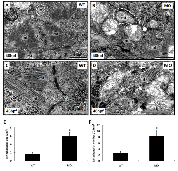
Fig. 7
Ultrastructural changes of mitochondria and myofibrils in the heart of tbx5-knockdown and wild-type (WT) embryos at 48 h post-fertilization (hpf) under TEM. A: Small mitochondria and well-formed desmosomes can be observed in WT embryos. B: Multiple swollen mitochondria can be observed within myocytes of tbx5 knockdown embryos. C: Well-organized myofibrils with a M line and Z disc can be observed in normal embryos. D: Disarrayed myofibrils appeared in tbx5 knockdown embryos while the M line and Z disc were unrecognizable. E-F: Both the size (E) and number (F) of mitochondria had significantly increased in tbx5 knockdown embryos. Data are presented as the mean � S.D. An asterisk indicates a significant difference (p < 0.05). mf, myofibril; mt, mitochondria; D, desmosome; ML, M-line; Z, Z-disc; white arrow, disarrayed myofibrils; MO, tbx5 knockdown embryos.