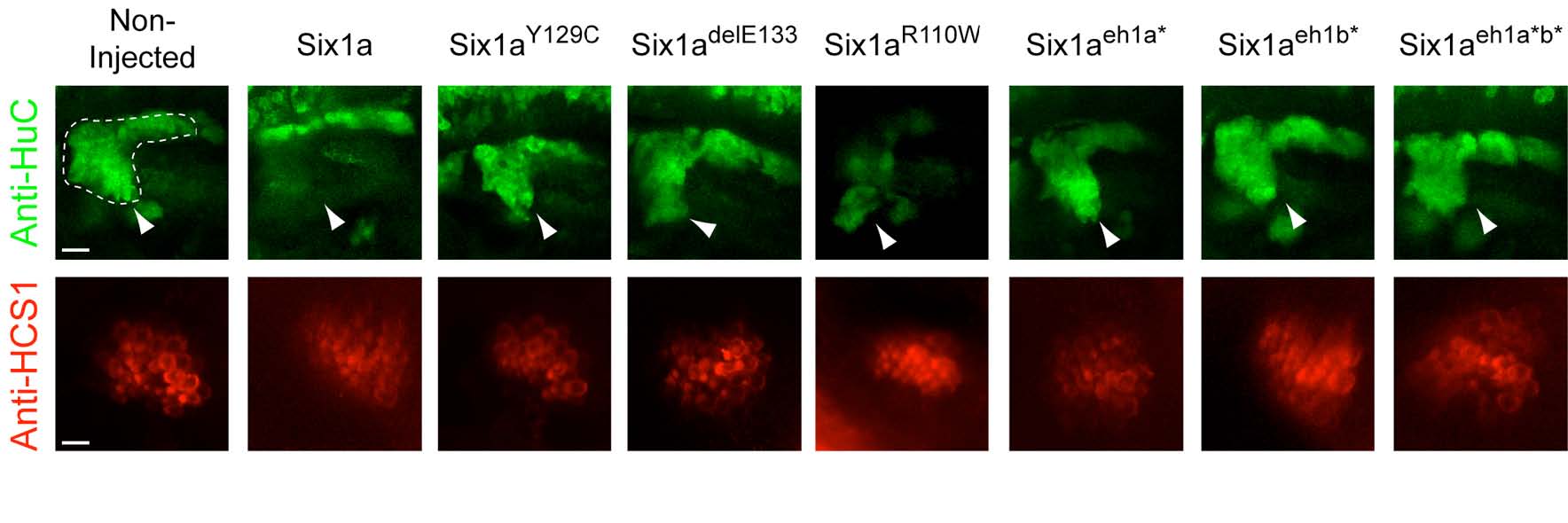
Fig. S2
Otic neurons and anterior macula hair cells in a 3 dpf developing zebrafish inner ear. Otic neurons in 3 dpf zebrafish larvae over-expressing mutant forms of Six1a have been detected by anti-HuC immunostaining (upper panels, in green; arrowheads and surrounded by a dotted line in the first panel) whereas hair cells of the anterior macula of 3 dfp zebrafish larvae over-expressing the same mutant forms of Six1a have been detected by anti-HCS-1 immunostaining (lower panels, in red). The upper panels are in a lateral orientation with a scale bar of 30 μm whereas the lower panels are ventral views with a scale car of 20 μm.
Reprinted from Developmental Biology, 357(1), Bricaud, O., and Collazo, A., Balancing cell numbers during organogenesis: Six1a differentially affects neurons and sensory hair cells in the inner ear, 191-201, Copyright (2011) with permission from Elsevier. Full text @ Dev. Biol.

