
Image
|
Figure Caption
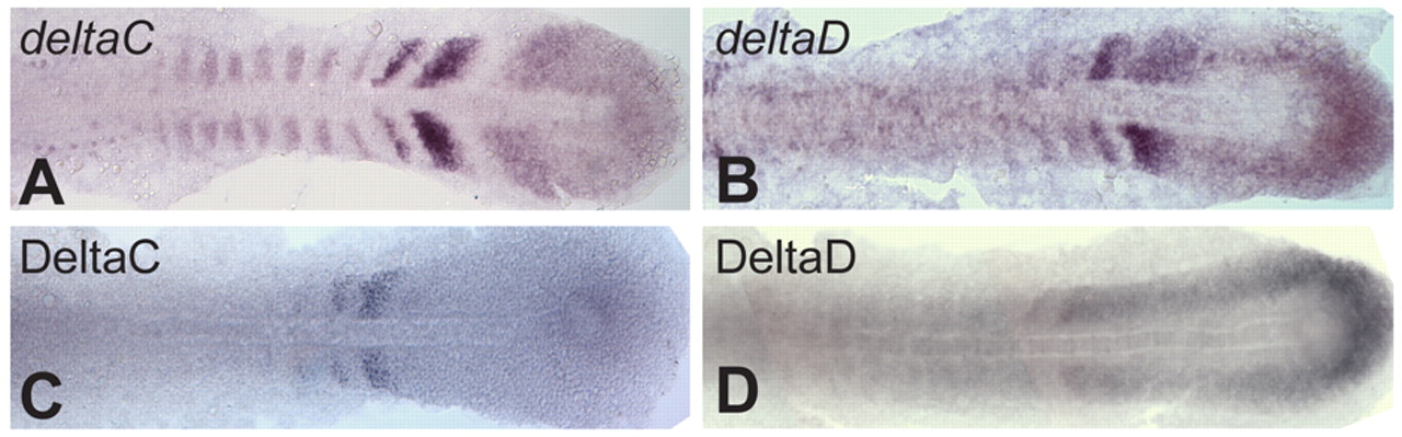
Fig. 4 ISH and antibody staining patterns compared in flat-mounted ~10-somite stage zebrafish embryos. (A,B) ISH for deltaC and deltaD. (C,D) Immunohistochemical staining of DeltaC with zdc2 and of DeltaD with zdd2.
Acknowledgments
This image is the copyrighted work of the attributed author or publisher, and
ZFIN has permission only to display this image to its users.
Additional permissions should be obtained from the applicable author or publisher of the image.
Full text @ Development