Fig. 1
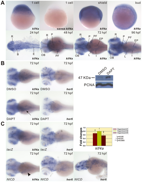
Developmental expression patterns of klf4a and the effect of Notch signaling on klf4a expression.
(A) Embryos from different developmental stages were hybridized with either antisense klf4a or sense klf4a RNA probes as indicated. Lateral view is shown for 1 cell, shield, and bud embryos. Dorsal view is shown for 24?96-hpf embryos. B, brain; E, epidermis; EP, exocrine pancreas; I, intestine, L, liver; OB, olfactory bulbs; P, pharynx; PF, pectoral fin; R, retina. (B) Enhanced klf4a expression in intestines (arrow) of DAPT-treated embryos is shown by in situ hybridization and Western blot analyses. Decreased her6 expression in the brain and intestines of DAPT-treated embryos is shown by in situ hybridization. Similar PCNA expression levels are shown in DMSO- and DAPT-treated embryos. (C) Decreased klf4a expression in intestines (arrowhead) of NICD-overexpressed embryos is shown by in situ hybridization and qPCR. Increased her6 expression in the brain and intestines of NICD-overexpressed embryos is shown by in situ hybridization. Error bars indicate the standard error. Student′s t-test was conducted to compare lacZ- and NICD-ectopically expressed embryos.

