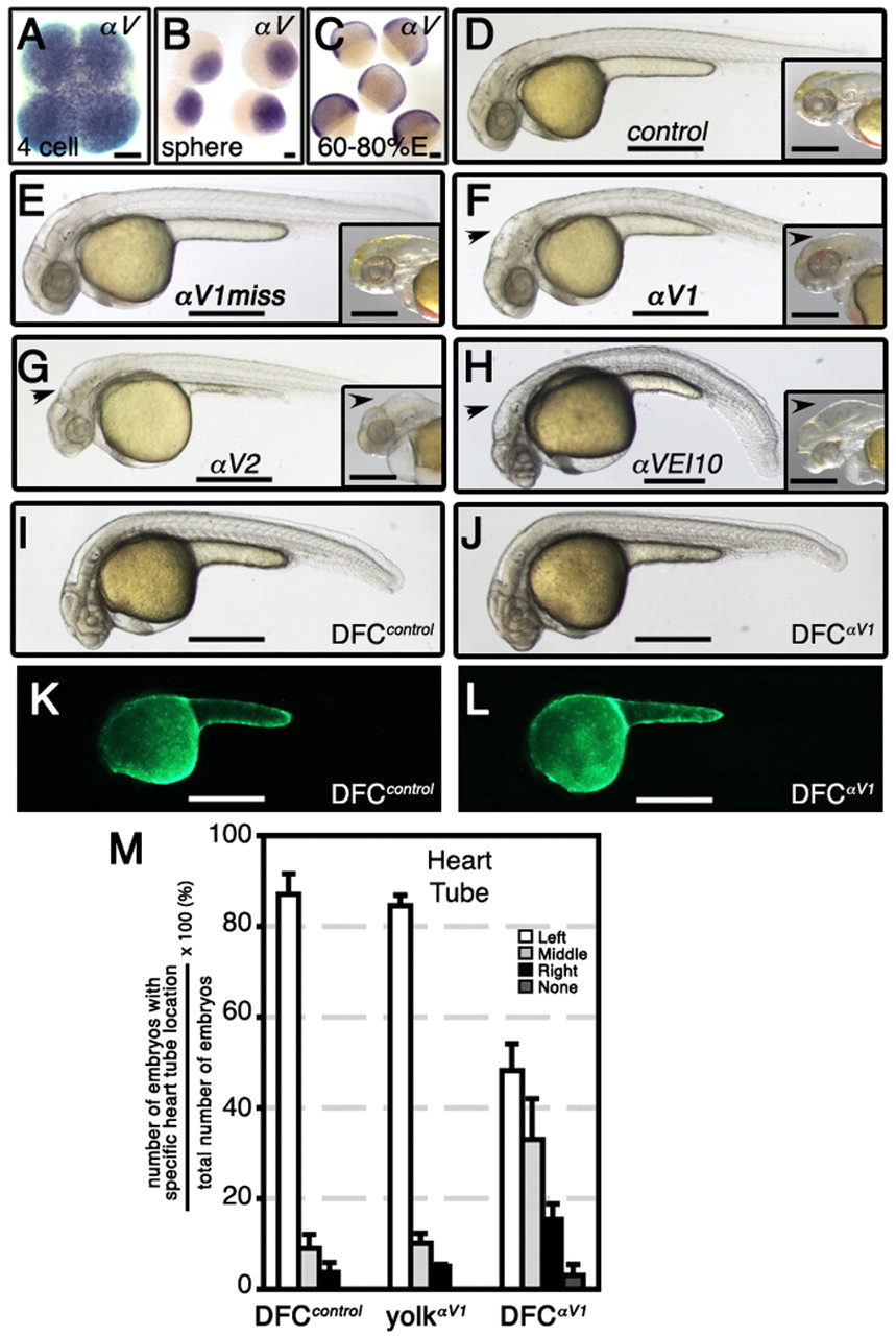
Fig. 2 Integrin αV mRNA is a maternal factor and its specific knockdown in DFCs alters heart tube asymmetry. (A-C) WISH analysis in wild-type zebrafish embryos shows maternal expression of αV at the 4-cell stage (A) and sphere stage (4 hpf; B), and zygotic expression at 60-80% epiboly (E). (D-L) Lateral views of live embryos at 32 hpf (D-H) or 28 hpf (I-L). When integrin αV morpholinos (MOs) were delivered at the 1- to 4-cell stage, morphants developed hydrocephaly in the fourth ventricle (black arrowheads) that was also associated with formation of abnormal cerebellum. Control (D) and αV1miss (E) morphants had wild-type head phenotype. Insets in panels D to H represent ∼55 hpf head phenotype of respective morphants. Note intracerebral bleeding (pink color behind eyes) at 55 hpf. When control or αV1 MOs were injected into yolk at mid-blastula stage (512-1000 cells), which targets DFCs specifically, these animals had wild-type phenotype (I,J). (K,L) Fluorescence images corresponding to I and J, revealing that MOs were exclusively present in the yolk cell. (M) Bar graph showing the effects of αV integrin loss-of-function specifically in DFCs on heart tube location. Data expressed are similar to those in Fig. 1E. Scale bars: 100 μm in A-C; 500 μm in D-L. See also Table S3 in the supplementary material.
Image
Figure Caption
Figure Data
Acknowledgments
This image is the copyrighted work of the attributed author or publisher, and
ZFIN has permission only to display this image to its users.
Additional permissions should be obtained from the applicable author or publisher of the image.
Full text @ Development

