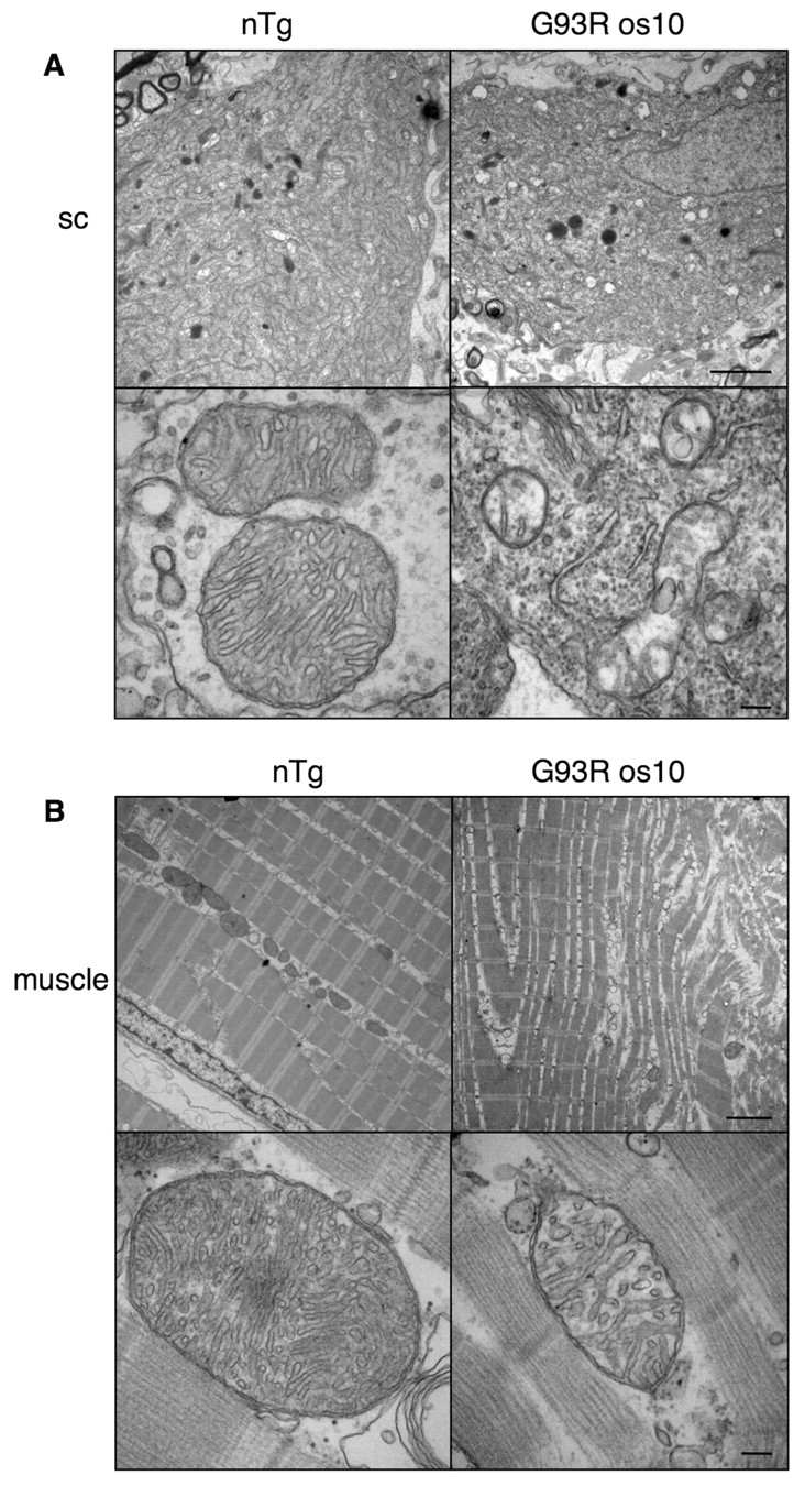
Fig. 6 Pathological changes were observed in G93R os10 spinal cord and muscle at the disease end-stage. (A) Electron microscopy of a spinal cord motoneuron from nTg sibling (left) and G93R os10 (right) adult end-stage fish. Numerous vacuolated mitochondria were observed in G93R os10 motoneurons (top) and were readily apparent at higher magnification (bottom). (B) Electron microscopy of trunk muscle from nTg sibling (left) and G93R os10 (right) fish also revealed severe abnormalities (top). The mitochondria in G93R os10 muscle also appeared to be affected, both in number and integrity, and cristae appeared to be reduced in number and lacked the normal organization (bottom). Bars, 2 μm (A,B; top panels); 150 nm (A,B; bottom panels).
Image
Figure Caption
Figure Data
Acknowledgments
This image is the copyrighted work of the attributed author or publisher, and
ZFIN has permission only to display this image to its users.
Additional permissions should be obtained from the applicable author or publisher of the image.
Permissions updated 10/14/2010.
Full text @ Dis. Model. Mech.

