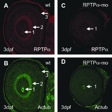
Fig. 4 Defects in retinal organization and differentiation in receptor protein-tyrosine phosphatase alpha morpholino (RPTPα-mo) -injected embryos. A,B: Oblique section of 3-days-postfertilization (dpf) -old wild-type (wt) embryo labeled with polyclonal anti-RPTPα antibody (AP5478; A) and anti-acetylated tubulin (B). Labeling is found in the optic nerve (arrow 1), the inner plexiform layer (arrow 2), and the outer plexiform layer (arrow 3). C,D: Oblique section of 3-dpf-old RPTPα-mo?injected embryo labeled with polyclonal anti-RPTPα antibody (AP5478; C), and anti-acetylated tubulin (Actub) antibody (D). Labeling of RPTPα and acetylated tubulin is found in the optic nerve (arrow 1), all other structures labeled in wild-type are not labeled in RPTPα-mo?injected embryos.
Image
Figure Caption
Figure Data
Acknowledgments
This image is the copyrighted work of the attributed author or publisher, and
ZFIN has permission only to display this image to its users.
Additional permissions should be obtained from the applicable author or publisher of the image.
Full text @ Dev. Dyn.

