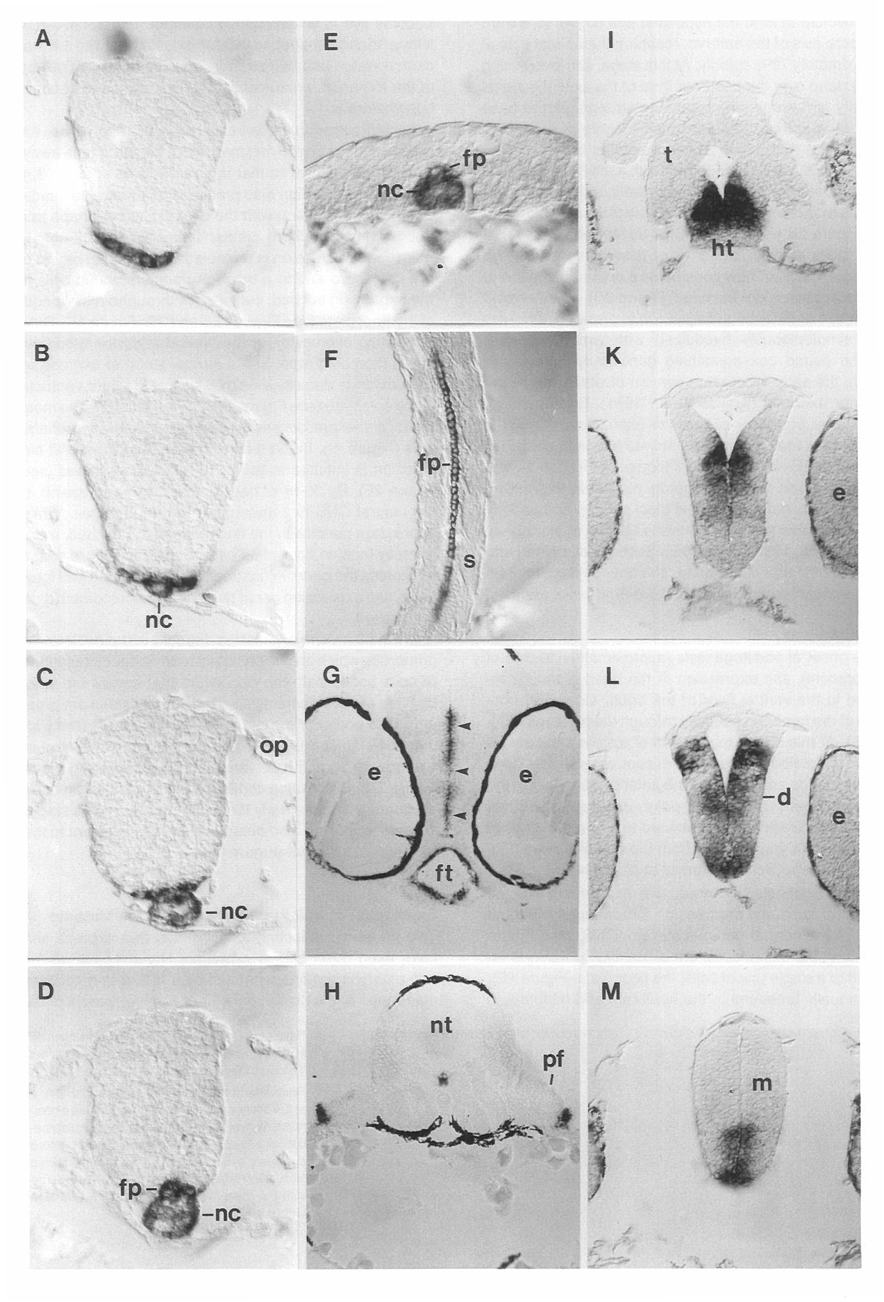
Fig. 4
Tissue Sections of Hybridized Embryos Showing shh Expression at Different Developmental Stages
(A-D) Successive transverse sections through the hindbrain of an embryo at the 5 somite stage showing shh expression. (A) Anterior hindbrain.
(B) Hindbrain at the anterior tip of the notochord. (C) Hindbrain at the position of the otic placode. (D) Posterior hindbrain.
(E) Transverse section through the tail of an embryo at the 5 somite stage.
(F) Horizontal section through the spinal cord of an embryo at 26 hr of development. Note the single row of floor plate cells that express shh.
(G and H) Transverse sections through a 48 hr zebrafish embryo. (G) Section through the diencephalon. shh expression is seen in the luminal cells of the foregut and in periventricular cells in the diencephalon (arrowheads). (H) Transverse section at the level of the pectoral fin buds.
(I-M) Serial transverse sections through the head of a 26 hr embryo.
Unless indicated, dorsal is to the top, Abbreviations: d, diencephalon; e, eye; fp, floor plate; ft, foregut; ht, hypothalamus; m, mesencephalon; nc, notochord; nt, neural tube; op, otoc placode; pf, pectoral fin; s, somite; and t, telencephalon.
Reprinted from Cell, 75(7), Krauss, S., Concordet, J.P., and Ingham, P.W., A functionally conserved homolog of the Drosophila segment polarity gene hh is expressed in tissues with polarizing activity in zebrafish embryos, 1431-1444, Copyright (1993) with permission from Elsevier. Full text @ Cell

